
The entopterygoid is a medial palatal bone in many ray-finned fishes, also called endopterygoid and historically mesopterygoid in relevant anatomical literature. It may form a thin lamina near the orbital floor or carry complex tooth surfaces in particular evolutionary lineages. An isolated archaeological plate can support identification only when its original contacts and preservation are understood; it cannot by itself establish fish numbers, specific diet or ancient human fishing practices.
This guide uses genuine comparative research on a Paleocene squirrelfish, Devonian lungfish, living characiforms and a correctly attributed historical 1866 perch skull drawing. Reed Basin and its worked numbers are fictional, not results from these studies. The discussion distinguishes measured bone anatomy, proposed biological functions and archaeological depositional histories.
Start with the 2021 palatal tooth evolution study, the Paleocene squirrelfish entopterygoid description and the Devonian lungfish palate investigation.


The entopterygoid evidence ladder
| Direct observation | Possible inference | Additional support required |
|---|---|---|
| Medial palate lamina and original contacts | Entopterygoid diagnosis | Taxon-matched skull and competing elements |
| Teeth, sockets or original toothless surface | Comparative dental character | Damage and species-specific references |
| Orbital-floor portion on a fossil scan | Possible orbital support | Associated skull anatomy; not direct eyesight |
| Recorded complete and fragmentary pieces | Sample condition and recovered concentration | Sediment denominators and recovery comparability |
| Bone in human settlement sediment | Possible fishing, trade or waste history | Positive site, transport and chronological evidence |
1. Begin with a medial palate bone, not a reconstructed fishery
The entopterygoid, also often named endopterygoid and historically mesopterygoid, lies within the palate of numerous ray-finned fishes. Its position is generally more medial than the ectopterygoid and may support portions of the mouth roof or orbital floor. It can be a flat lamina, a thickened tooth-bearing plate or a differently shaped structure according to taxon. Archaeological fragments sometimes retain its contacts or dental features, but a single piece does not reveal one fish’s age, prey or the number of fish people caught.
The first useful field record describes the bone’s thickness, original and broken margins, any tooth-bearing face and possible contact with the palatine or metapterygoid. A suggested species comes later, after actual comparative fish references. Human processing, trade and deposit chronology are separate questions. This sequence prevents a thin plate from becoming an elaborate but unsupported historical narrative.
2. Locate it medially within the palatal arch
The medial roof of the mouth in many teleosts includes entopterygoid and adjacent bones, while the ectopterygoid tends to form a more lateral palatal region. Posterior contacts may involve metapterygoid, quadrate and other suspensorial components. The precise layout differs among species. A plate recovered without original context can resemble a broad cranial wall or opercular fragment, so one frontal or lateral sketch seldom supports a firm anatomical identification.
Students should label entopterygoid, ectopterygoid, palatine, metapterygoid and quadrate on an identified reference skull. They then remove the neighbouring bones and state which diagnostic surfaces are lost when the element becomes isolated. The answer should be a list of observed contact characters rather than a generic shape comparison.
3. Recognise historical mesopterygoid terminology
Older fish osteological works regularly use mesopterygoid for a bone now called endopterygoid or entopterygoid in appropriate contexts. The terms should be mapped carefully by the original drawing and taxon, rather than replaced mechanically in every historical publication. Richard Owen’s nineteenth-century comparative figures used particular numbering and descriptive relationships that need to be read in their historical nomenclatural frame.
Ask learners to create a terminology crosswalk containing the original name, original author’s fish, anatomical number or contact, and proposed current counterpart. Preserve the old label in the archive. A transparent crosswalk allows future researchers to reassess a homology rather than assume old and new words always refer to identical material.
4. Read the actual 1866 perch plate correctly
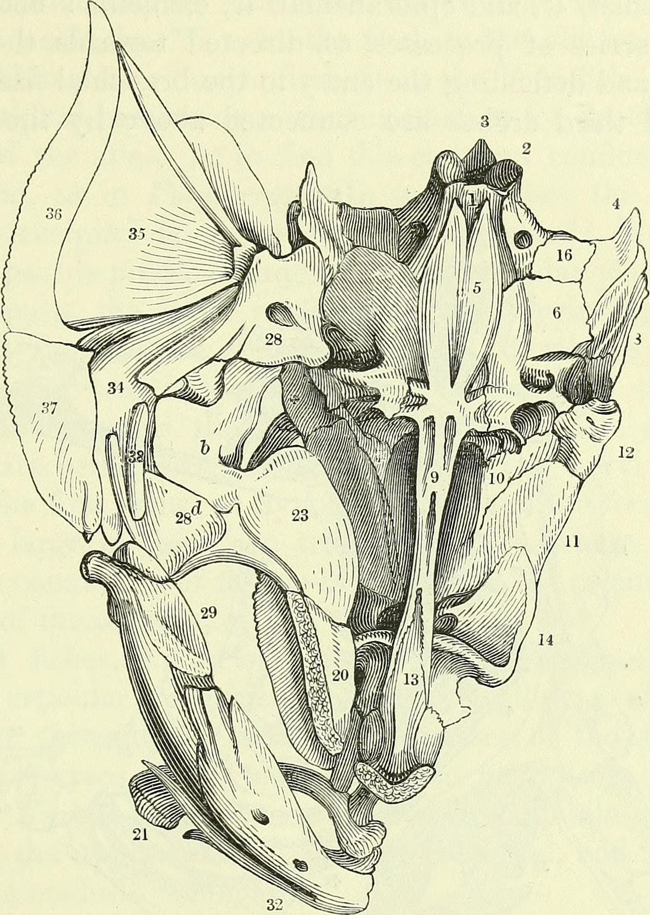
The featured public-domain image is from Richard Owen’s *On the Anatomy of Vertebrates* (1866). It illustrates the base of a European perch skull and part of the mandibular and opercular apparatus, with bone 23 identified in the accompanying text as entopterygoid and bone 20 as palatine. The illustration is a historic reference drawing, not a micrograph of a modern specimen or a fish excavated from the fictional Marsh Quarry described later.
Students should locate 23 and 20 and confirm what each label means in the accompanying original text. The diagram’s historical perspective and unscaled reproduction require care: an illustrated proportion should not be copied into a numerical fish-size model without specimen data. Accurate captions describe both the source and its limitations.
5. Distinguish entopterygoid from ectopterygoid
Ectopterygoid is often a more lateral palatal element, whereas entopterygoid sits medially or dorsomedially and may spread into the orbital floor. In some taxa the two bones overlap and one contact is broad, making fracture lines hard to interpret after burial. A dental surface alone cannot always identify which bone contributed it. Comparative whole-skull evidence matters more than a convenient position on a two-dimensional diagram.
Students should examine an identified reference in lateral and medial views and mark original bone boundaries. If a fossil lacks the separating contact, a broad palatal-fragment identification may be the correct result. An ambiguous piece must not be entered simultaneously into both accepted skeletal categories simply because both interpretations remain possible.
6. Distinguish it from metapterygoid
The metapterygoid arises in a different relationship to the palatoquadrate and often occupies a more posterior suspensorial position. Entopterygoid may overlap or meet it along the palatal and orbital region. Their outlines can both be plate-like, and their original margins may be damaged at the contact, especially in flattened fossils. Developmental descriptions and osteological studies are essential for deciding whether a loose fragment came from the medial palate or the posterior suspensorium.
Have students compare a securely identified pair from one reference fish. They record which original margin belongs to the entopterygoid and which to the metapterygoid. If the fossil preserves only a central patch, the analysis should retain anatomical uncertainty instead of deriving a confident species identification.
7. Explain the orbital-floor relationship without exaggeration
In several fish lineages the entopterygoid contributes a thin lamina near the underside of the orbit, while other palatal and neurocranial bones share this region. The anatomical role is not a universal uniform plate supporting the eye in exactly the same way in every fish. Bone extent and adjacent contacts can change in evolution. A fossil entopterygoid may show a thin expansion but cannot alone reconstruct the eye’s size or sensory capability.
Students should draw the orbit, entopterygoid lamina and neighbouring palatine in a documented skull. They should distinguish preserved hard tissue from missing soft tissue and eye anatomy. This guards against false claims that one plate width directly measures how a fish saw prey.
8. The earliest Paleocene squirrelfish offers a detailed fossil comparison
A 2023 study by Andrews and colleagues described an early Paleocene squirrelfish relative and documented a fractured entopterygoid still associated with palatine, ectopterygoid and metapterygoid. The described lamina curved medially to help define the floor of the orbital cavity. The fossil also illustrates a research limitation: tomography did not show obvious entopterygoid dentition in the material available. Non-detection under those conditions is not a global evolutionary rule about all squirrelfishes.
Students should distinguish what the scan revealed from what the authors inferred about missing margins. They should identify the fossil’s geological age and not turn it into a human archaeological fish bone. The value of the paper lies in its precise preservation-aware description of contacts.
9. Interpret ‘no teeth in the scan’ narrowly
A fossil paper may report that no obvious dentition appears on an entopterygoid under a particular tomographic resolution. That is a statement about the observed specimen and detection method. Fine teeth could be absent biologically, weathered away, obscured by matrix or below the scan’s resolution. The strongest claim must reflect the observation rather than translate a non-detection into an absolute species-family rule without suitable reference.
Ask learners to write three alternative explanations for a scan with no visible teeth and identify one test for each. An indeterminate dental condition is a legitimate scientific category when preservation is insufficient.
10. The 2021 characiform study demonstrates variable teeth
Jandzik and Stock surveyed tooth-bearing bones in characiform fishes and analysed phylogenetic changes in dental distribution. Their study identified multiple independent gains or re-gains of entopterygoid teeth among the sampled lineages. This contradicts any simple rule that a medial palatal bone is universally toothless or always toothed. The ancestral-state models infer evolutionary histories from reference character distributions; they are not direct observations of individual ancient fishes.
Students should keep two layers of evidence distinct: actual teeth or absence in identified species, and the inferred evolutionary changes mapped onto a tree. A phylogenetic estimate cannot by itself identify the taxon of one archaeological tooth plate with missing margins.
11. Characiformes and Cypriniformes should not be merged
Characiform fishes may carry palatal teeth in several places, including ectopterygoid and entopterygoid in certain taxa. Cypriniform fishes such as zebrafish and carp have much more restricted tooth locations, famously concentrating functional teeth in the lower pharynx. This broad contrast is based on comparative biology, but species variation and evolutionary history still matter. Not every characiform species displays the same palatal dentition.
Have students compare documented dental maps of a Mexican tetra and a toothed-palate characiform such as *Hoplias*. They should locate the original bones before comparing teeth. A loose crown from a river deposit cannot be assigned to entopterygoid simply because a characiform source is nearby.
12. Learn what developmental potential really establishes
The 2021 study experimentally altered ectodysplasin signalling in model characiform fish and observed an expanded distribution of teeth. This supports a conclusion about the developmental capacity for tooth formation in that lineage under experimental conditions. It does not prove that a historic fish possessed laboratory-induced teeth or that each tooth recovered archaeologically records a genetic mutation.
Students should distinguish experimental evidence from fossil evidence and avoid inventing causal genes for a single old bone. The study informs possible evolutionary mechanisms while the archaeological specimen remains an observation of a particular physical object.
13. Devonian lungfish reveal another tooth-bearing pattern
Research into the early evolution of lungfish hard-food processing describes a large entopterygoid bearing patterned conical teeth and a finer denticulate lamina, alongside dermopalatine and ectopterygoid regions. The fossil illustrates that palatal dentition can be morphologically complex in deep-time sarcopterygians. That architecture is not a species diagnosis for a modern teleost in an archaeological settlement.
Ask learners to mark which lamina bears dense fine denticles and which has larger rows, using the original paper’s actual descriptions. Then they should explain why the evolutionary and taxonomic context prevents direct transfer of its diet to an unrelated fish’s detached entopterygoid.
14. A tooth plate may participate in hard-food processing
Strongly developed palatal teeth may help secure or process hard prey in some fishes, but a bone’s tooth pattern does not reveal an individual last meal. Functional hypotheses can be tested against modern biomechanics, wear and ecological reference, while fossil interpretations require cautious comparisons. A dentigerous entopterygoid in a Devonian lungfish cannot be used as universal evidence that all toothed palatal plates broke shells.
Ask students to list two distinct potential mechanical functions compatible with a broad tooth plate and the additional observations needed to discriminate them. The final report should describe observable teeth and plausible feeding capacity without naming a particular historical prey.
15. The Triassic Marcopoloichthys figure illustrates a medial contact
Gloria Arratia’s 2022 reconstruction of the Triassic *Marcopoloichthys furreri* labels ent for entopterygoid, ect for ectopterygoid and par for parasphenoid. The two illustrated head positions convey an interpretation based on several fossil specimens, not direct observation of a living Triassic feeding motion. The spatial relationship of entopterygoid and other palatal elements is valuable for anatomical comparison even though the figures are reconstruction.
Students should compare the actual source diagram with the 1866 perch plate. They should state what is shared about skeletal location while acknowledging enormous geological and phylogenetic differences. A reconstructed contact cannot replace direct observation of a fragment’s original articular surface.
16. A historical plate can contain outdated names
Richard Owen’s 1866 terminology and modern developmental bone names cannot be assumed interchangeable at face value, especially when the original figure and text divide the pterygoid region differently. A historical drawing is valuable for locating a region but may omit fine anatomical contacts or fuse interpretations in ways later scholarship revised. Citing the original source and explaining its limits is better than silently upgrading every old label.
Students create a comparison note with the original numerical bone labels and current terms as used in an appropriate source fish. They should not erase the historic name from the permanent specimen metadata.
17. Do not treat entopterygoid as a generic ‘fish eye bone’
Although some entopterygoid laminae contribute beneath the orbit, many other elements surround the fish eye, including frontal, infraorbital and lateral ethmoid components. Their species-specific positions differ. An isolated plate found near orbital material in a burial is not necessarily entopterygoid based on spatial proximity after disarticulation. Correct identity rests on preserved original contacts rather than the nearest excavated object.
Have learners sort a group of labelled modern skull bones before comparing them with a set of isolated fragments. They should preserve an indeterminate cranial-plate category when necessary to prevent one uncertain object from becoming a falsely diagnostic species record.
18. Record teeth, sockets and damaged tooth rows separately
Teeth can be worn, replaced, lost or broken from palatal surfaces, leaving small sockets or eroded bases. A thin lamina may also be partially dissolved, making the original tooth distribution hard to reconstruct. If one fossil has obvious teeth and another lacks visible crowns, they should not automatically be coded as different species without considering preservation. Both tooth and supporting bone need anatomical description.
Students should record whether crowns, sockets and original margins are present and whether the surface is intact enough for biological absence to be diagnosed. A fragment lacking its tooth-bearing edge is unsuitable for a confident toothless category.
19. Morphological variation includes life stages
Palatal bones can grow and change proportions as a fish matures, and dental patterns may differ with age or tooth replacement. A juvenile entopterygoid can have a less conspicuous lamina or articular contact than an adult reference. Anatomical comparisons should therefore use a size-appropriate modern series where possible and avoid assigning unusual juvenile shapes to a new fossil species without evidence.
Students should compare juvenile and adult identified reference specimens, noting which contacts are already ossified and which surfaces are incomplete. Exact age-at-death cannot be inferred from one lamina without a validated age model.
20. Take medial and lateral orientation seriously
An entopterygoid may be broad but thin, and viewing it from the opposite surface can change the apparent curvature, dental margin and overlap with neighbouring bones. Flattening during burial may distort these characters further. A scientific drawing should state left or right side, medial or lateral view, scale and whether it represents actual fossil margins or restored anatomy.
Have students rotate a specimen in multi-view photographs and identify original articular surfaces. A flat image alone may provide sufficient evidence for a broad element but not a narrow species claim.
21. Careful tomography may improve diagnosis but cannot restore missing bone
Micro-CT can reveal internal contacts, tooth bases and buried plate portions when mineral contrast permits. It is valuable for rare fossils because it may reduce the need for destructive preparation. But resolution, matrix density and reconstruction choices introduce limits. A scan does not manufacture the original outline where erosion removed it, nor can it establish toothlessness if tiny crowns fall below detection.
Students should compare the documented Paleocene squirrelfish fossil scan with a hypothetical damaged archaeological plate. They identify what information imaging could add and which uncertainty would remain even with excellent resolution.
22. Taxonomic resolution should match actual preserved characters
Entopterygoid morphology may contain species-level differences for some groups, but shared shapes and incomplete local reference collections limit interpretation. A medieval fish bone could be readily identified to a broad teleost category while resisting a confident genus assignment. The correct rank is the finest one supported by direct characters and exclusions of plausible alternatives.
Ask learners to write which preserved contact supports each suggested rank. If only a shared lamina is present, record palatal element or entopterygoid-type fragment rather than the fish whose image looks most familiar.
23. Measurement of a lamina needs original landmarks
Height, width, overlap length and tooth-row spacing can be measured on complete reference entopterygoids, but each measurement requires a defined axis and original preserved margins. A thin lamina with a fractured end gives a partial dimension, not the full input intended by a species-calibrated relationship. Because fish skull proportions vary, a body-size formula from a different bone cannot be substituted.
Students should draw two measurement lines on a known modern specimen and attempt to reproduce them. If one endpoint is missing in the fossil, record the reliable partial measurement and withhold the complete estimate.
24. A fictional Reed Basin geometry comparison
In the entirely invented Reed Basin teaching example, a complete entopterygoid lamina A measures 8.0 mm along a specified original long axis and 4.0 mm along an orthogonal width, giving a ratio of 2.0. Reference B is 10.0 mm by 5.0 mm, also 2.0. The shared ratio does not indicate identical taxa, absolute fish length, feeding function or geological period. These are made-up demonstration measurements and not values from the Owen or squirrelfish illustrations.
Students should keep original measurements beside the dimensionless ratio and state the need for independent modern references. Numerical similarity is descriptive, not proof of one biological identity.
25. A cracked lamina cannot yield a whole-bone ratio
A plate fractured through its original margin may still be measured precisely as a remnant, but the result differs from the complete-bone length or width used in a calibration. Extrapolating by copying a corresponding margin from a different fish species adds unmeasured assumptions. A damaged original boundary should remain missing in the dataset rather than turn into a false decimal.
Provide a fictional specimen with original width but lost long-axis endpoint. Learners record width and flag the whole-bone ratio as unavailable, while retaining the specimen for other diagnostic characters.
Reed Basin: wholly fictional original dimensions
| Toy plate | Original long axis | Original width | Long / wide |
|---|---|---|---|
| A | 8.0 mm | 4.0 mm | 2.00 |
| B | 10.0 mm | 5.0 mm | 2.00 |
| Damaged C | Original edge missing | 4.0 mm | Unmeasurable |
26. Count fragments and original bones as distinct units
A fish may have paired entopterygoids and one may break into multiple identifiable shards. The number of physical pieces is not the number of original animals. Archaeological catalogues need separate fields for complete elements, diagnostic fragments, refits, unidentifiable pieces and confidence. An apparent increase in fragments between two layers can reflect breakage rather than a larger source fish population.
Students should refit a pretend plate from four pieces and document all observations. A fish count derived by assigning one animal per piece would be demonstrably wrong.
27. Minimum-individual estimates need secure sidedness
Within a coherent, narrowly defined archaeological context, independently identified same-side entopterygoids from one species may help constrain a conservative minimum number of fish. A group of unsided shards cannot simply be added as animals. Nor does one biological species automatically follow from one shared palate plate morphology. A valid minimum estimate depends on taxonomic identity, anatomical repetition and context boundaries.
Compare three secure left bones with sixteen small unplaced fragments. Students should distinguish minimum individuals from the total observed specimen count and state which assumptions make any estimate possible.
28. Recovery methods can create apparent absences
Entopterygoid plates may be thin and fracture into small pieces that escape coarse sieves. Differences in wet processing, screen fraction, sediment mass and sorter experience also affect detection. A site with few diagnosed entopterygoids cannot be said to lack fish with such bones if the recovery procedure was unsuitable. Negative archaeological evidence is conditional on the probability of detection.
Students should audit two excavation logs and consider whether fine-screened archived samples could be compared. The correct inference may be that available material is not sufficiently standardised to support an abundance trend.
29. A fictional Reed Basin fragment comparison
Imagine an older Reed Basin layer with fifty-two confidently classified entopterygoid objects, thirty-two intact and twenty fragmented, from 2.6 kg of dry sediment. A younger layer has seventy-eight objects, twenty-six intact and fifty-two fragmented, from 6.5 kg. The observed total rates are 20 and 12 objects per kilogram. The younger raw tally is higher, but its rate per processed sediment is lower.
Students must calculate both and retain the original count units. Reed Basin and every number are fictional, not findings from a real archaeological fishery. A simple per-mass ratio is not a biological population or annual catch estimate.
30. Reed Basin’s intact fraction gives a different clue
In the imaginary older layer, intact specimens account for 32/52, or about 61.5 percent. In the younger, 26/78, or one-third. Complete specimens per kilogram therefore change from about 12.31 to 4.0. This difference may be caused partly by transport, sampling or burial damage, making a biological source interpretation uncertain without a preservation audit.
Have students graph both total and intact object rates, identify the different numerators and request independent measurements of abrasion or sediment energy. A reliable report can make a precise descriptive statement without claiming to know the unique ecological cause.
Reed Basin: fictional bone-count audit
| Toy layer | Complete | Fragments | All diagnosed objects | Dry sediment | Objects per kg | Intact share |
|---|---|---|---|---|---|---|
| Older | 32 | 20 | 52 | 2.6 kg | 20 | 61.5% |
| Younger | 26 | 52 | 78 | 6.5 kg | 12 | 33.3% |
31. A fish plate near people can come from several pathways
An entopterygoid can enter a sedimentary layer through natural fish mortality, predator feeding, human processing, trade in fish products or later flood transport. A marine fish palate recovered inland may be imported rather than evidence of an ancient sea in the settlement. Human proximity and skeletal identity are separate observations. A tooth-bearing plate beside a hearth does not independently establish that the fish was cooked there.
Students should develop at least two formation histories for an imaginary deposited palate bone and name one field observation that would help discriminate them. Cut marks, anatomical assemblage, burning pattern, predator modification and sediment layers may all matter.
32. Age estimates must not be invented from palate morphology
Entopterygoid outline and tooth distribution usually do not provide a direct calendar year for the fish’s death or its sedimentary burial. Biological age, geological age and human archaeological deposition are different variables. A geological fossil from the Paleocene or Devonian cannot be described as food remains from a later human community. An archaeological bone may also be older than the layer where it was reworked.
Students should prepare three time labels for each source: taxon geological period, individual biological lifespan where known, and depositional age. Their bibliography and captions should retain these distinctions.
33. Preserve specimens and document scientific images
Rare plate-like skull bones may be lost or damaged by aggressive cleaning, drilling or resection. Any destructive analysis needs authorised custodial permission and a question-specific gain. A public-domain historical diagram can be used in teaching but must retain its author, date, original bone number and limitation. Original scientific fossil reconstructions should be distinguished from photographed physical specimens and from fictional sample calculations.
Students should create a specimen custody and image attribution sheet. A second reviewer should be able to repeat the identification and count without guessing the source of the plate or the meaning of its numbering.
34. Conclude at the strongest supported level
The invented Reed Basin data describe changing recovered counts and completeness, not a real fish population trend. Genuine studies show that entopterygoid dentition can vary substantially and that some fossil plates contribute to orbital-floor reconstruction. These observations do not permit the exact prey, individual fish age or human catch technique to be inferred from one detached medial palate bone. Each stronger claim requires independent evidence.
The gold standard is a transparent anatomical diagnosis, well-defined measurements, original observation counts, preservation assessment, appropriate comparative taxonomy and a source-history explanation clearly labelled as testable rather than certain.
Twenty-six applied research and archaeology cases
The exercises are fictional decision problems, not additional real discoveries. They test identification, measurement and inference independently.
1. Owen’s number 23
A student labels number 23 in the 1866 perch plate as a metapterygoid. The group checks Owen’s accompanying description and corrects it to entopterygoid within the historical nomenclature.
2. A modern adult plate with ancient provenance
A public-domain 1866 drawing is presented as an excavated medieval fish fragment. Students restore actual source, date and scientific purpose and separate it from Reed Basin’s fictional calculations.
3. Entopterygoid confused with ectopterygoid
A tooth-bearing fragment is called ectopterygoid without a preserved outer palatal contact. Learners compare medial and lateral positions before selecting a supportable diagnosis.
4. The missing metapterygoid contact
A plate is fractured at its posterior margin. Students preserve a broad palatal classification rather than infer a unique species from an unobservable metapterygoid contact.
5. Paleocene squirrelfish toothless?
A damaged fossil scan shows no clear entopterygoid teeth. The class distinguishes non-detection from biological absence and lists image-resolution and preservation limitations.
6. A Devonian lungfish used as a modern fish key
An old lungfish with elaborate tooth rows is treated as the species reference for a teleost archaeological fragment. Students explain why shared bone names do not make taxon-specific diagnoses portable.
7. The thirteen rows in a paper
Published lungfish teeth are counted and a student assumes the same number must occur in all fishes. Learners state the species and geological context before using the finding.
8. The 2021 tooth-evolution tree
Ancestral-state inference is presented as a directly photographed ancestor. Students label the phylogenetic reconstruction separately from observed living specimens and fossil evidence.
9. The wrong historical mesopterygoid label
Two museum catalogues use different pterygoid terms. Students compare original figures and preserve a terminology crosswalk rather than silently merge entries.
10. A tooth from an unknown bone
A loose crown is assigned to entopterygoid because the selected article is about that bone. Learners inspect supporting morphology and retain an unknown tooth location where necessary.
11. The orbital floor overclaim
One thin plate is used to infer fish eye size and visual acuity. The class identifies missing skull and soft-tissue evidence before making an ecological claim.
12. The mirrored reference view
A medial plate is compared with a reflected lateral image. Students align original sides and contact facets to prevent false species differentiation.
13. A juvenile plate’s outline
A small fish’s plate seems unlike the adult specimen in a guide. Learners review developmental variation before naming another taxon.
14. A socket remains without a crown
A worn margin is coded as naturally toothless. Students record tooth sockets and preservation before inferring dental absence.
15. A falsely precise 2.0 ratio
A toy specimen measures 8 by 4 mm and gives 2.0. Students state why it is geometry, not a real fish-length calibration.
16. Two sizes, identical proportions
A second toy plate of 10 by 5 mm also yields 2.0. The group explains why identical ratios do not imply the same species or absolute size.
17. A fractured original margin
A fossil’s width survives but not its full length. Students preserve the width while withholding a whole-bone ratio that requires the missing endpoint.
18. One bone split into several objects
A single entopterygoid fragments into four pieces during processing. Learners record physical fragments and possible refits instead of four fish.
19. Three same-sided references
A closed context has three well-identified left entopterygoids of one taxon. Students discuss a minimum-individual constraint and why unsided shards do not increase it mechanically.
20. Reed Basin’s raw count reversal
The younger fictional layer has more raw pieces but fewer per kilogram. Students compute 20 versus 12 and distinguish sampled sediment from fish abundance.
21. Intact share as preservation signal
The intact fraction changes from about 61.5% to 33.3%. Learners investigate abrasion and processing before attributing it to a biological population trend.
22. An excavation sieve becomes finer
A later excavation recovers more thin palatal plates because the mesh changed. Students assess method bias before claiming new fish arrived.
23. A fossil near a hearth
An entopterygoid is darkened near food remains. Learners distinguish cooking from secondary burning and reworked sediment using independent context.
24. Marine fish transported inland
A marine palatal plate is found in a river settlement. The class examines trade or transported fish products rather than an invented former local sea.
25. A wrong body-size formula
A dentary-height equation is supplied with entopterygoid width. Students reject the skeletal input mismatch despite identical units.
26. The final inference audit
One plate is said to prove species, prey, capture season, fish population and exact settlement year. Students preserve only direct anatomy and supported context, specifying tests for other claims.
Teaching sequence: three scientific sessions
- Session 1 — recognise entopterygoid versus ectopterygoid and metapterygoid, and read Owen’s original bone labels with a terminology crosswalk.
- Session 2 — examine observed orbital-floor laminae, characiform teeth evolution and fossil lungfish dentition, distinguishing reconstructed biology from source-specific evidence.
- Session 3 — work fictional geometry and sediment denominators, evaluate fragments, recovery and depositional agents, then write an evidence-bounded historical conclusion.
- Student submission — annotated comparative plate, preserved original landmark record, taxonomic confidence, count and denominator table, source attribution and an independent follow-up test.
Research-grade assessment rubric
| Question | Adequate evidence | Incorrect shortcut |
|---|---|---|
| What element survived? | Medial contacts, orientation and preservation recorded | Any palate plate named entopterygoid |
| What teeth were present? | Actual crowns, sockets or biological absence confirmed | Damage interpreted as inherited toothlessness |
| Can function be inferred? | Source-specific skull and relevant living ecology | Palate lamina used to infer eye acuity or a last meal |
| Can size be calculated? | Complete genuine landmarks and validated model | Ratio of toy dimensions called fish length |
| How many fishes? | Paired elements, refits and sample mass distinguished | Each fragment called one caught fish |
| What happened at site? | Predator, natural death, food use, trade and dating tested | One plate equated to one meal or year |
| Is the illustration accurate? | Owen 1866 source and bone number properly disclosed | Historical drawing passed off as excavated fossil |
Frequently asked questions
What is a fish entopterygoid?
A medial palatal bone in many fishes, also called endopterygoid and historically mesopterygoid in appropriate contexts.
How does it differ from ectopterygoid?
They generally occupy different medial and lateral palatal regions, though preserved contacts and taxonomic reference determine precise identification.
Does the entopterygoid always have teeth?
No. Dentition varies widely across fish lineages, including evolutionary gains and losses in characiforms.
Does an entopterygoid support the fish eye?
In some taxa its lamina contributes near the floor of the orbit, but it is not the sole eye-supporting structure in all fishes.
What does number 23 mean in the 1866 drawing?
The historical plate’s accompanying text identifies number 23 as entopterygoid in the reference perch.
Can it indicate ancient fish body length?
Only with a real validated taxon-specific model using intact original measurement landmarks.
Do palatal teeth reveal exactly what a fish ate?
No. They may inform feeding-function hypotheses but not a direct last-meal record.
Does one fragment mean one fish?
No. One paired skull bone can produce multiple fragments, so specimen counts and original individuals differ.
Are Reed Basin’s measurements real excavation data?
No. Reed Basin, its object totals and geometry are entirely fictional teaching examples.
How can a source history become more defensible?
By integrating anatomical diagnosis with actual stratigraphy, recovery methods, taphonomy, transport, processing and historical evidence.
Primary research and connected eduKate Gold Standard articles
- Owen (1866), original European perch entopterygoid number 23 plate
- Australian Museum, striped sea bass skull and labelled endopterygoid
- Jandzik & Stock (2021), entopterygoid teeth losses and re-gains in characiform fishes
- Early Paleocene squirrelfish entopterygoid and orbital floor (2023)
- Early lungfish hard-food processing and entopterygoid laminae
- Arratia (2022), Triassic Marcopoloichthys palatal contacts
- Evolution and development of fish jaw skeleton
- Palatoquadrate bone homologies and development
- Keivany (2015), comparative fish suspensorial and palate terminology
- Colley & Brownlee, anatomical recording conventions for fish remains
- Teaching Archaeological Fish Ectopterygoid Analysis
- Teaching Archaeological Fish Metapterygoid Analysis
- Teaching Archaeological Fish Palatine Analysis
- Gold Standard of Archaeology
- Gold Standard Master Hub
The gold standard of teaching entopterygoid analysis is a learner who identifies the medial palatal structure from real original contacts, distinguishes scientific observations from phylogenetic and biomechanical inference, and states what the sampled fragments and deposit can support. Even where fish species or ancient activity remains uncertain, a carefully documented palate bone improves an archaeological inventory.