
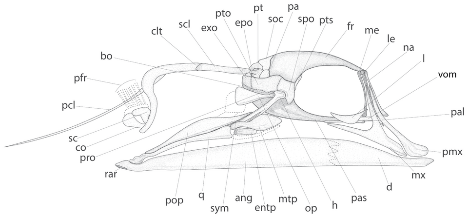
The metapterygoid is a bone of the posterior palatoquadrate and suspensorial region in many ray-finned fishes. It can help archaeologists describe cranial remains and compare taxa, especially when original contacts with the quadrate, palate or hyomandibula survive. But its outline varies with fish group and developmental stage. One isolated plate is not a fish census, a bite-force reading or proof of any particular human fishing practice.
This scientific teaching guide explains the metapterygoid–quadrate fenestra, the separate pseudobranchial-related foramen, growth-stage ossification, competing catfish homologies and a genuine 2026 Eocene fish comparison. The worked Brook Channel examples are fictional. The header figure is an interpretative reconstruction of a geological fossil, not a photographed archaeological sample or evidence for an ancient human community.
Core research includes the ZFIN anatomical definition, developmental osteology of Moenkhausia and the 2026 Eocene stylephorid paper.


Metapterygoid evidence overview
| Direct evidence | What it may tell us | What remains unsupported |
|---|---|---|
| Posterior palatoquadrate plate with correct contacts | Metapterygoid identification | Universal species or one original fish |
| Original fenestra margins | Comparative opening shape within a taxon | Unique function across all fishes |
| Verified pseudobranchial-related foramen | Specific developmental anatomical character | Exactly how one fish fed |
| Measured original bone height and width | Defined geometric ratio | Fish body length without a modern model |
| Sediment object counts | Sampled fossil inventory | Historic fish catches or human diet |
1. The metapterygoid is a particular skull bone, not a fossil fish count
The metapterygoid is a bone associated with the posterior palatoquadrate region and the suspensorium of many ray-finned fishes. It may form a broad plate or a more specialised configuration between the quadrate, hyomandibular region and palate. Its size and contacts vary substantially across fish families and growth stages. An archaeologist can sometimes recognise it from isolated articular or plate fragments, but one element does not independently establish the number of original fishes, feeding force or a named human activity.
Ask learners to inspect the labelled modern or fossil skull reference before naming a loose element. They should move from observed bone surface to source-taxon comparison and only then to depositional context. A precise anatomical description is useful even if the fish species remains unknown. Jumping from a plate-shaped fossil to a history of fishing is an evidential error.
2. Locate the bone behind the palate and above the quadrate
In many teleosts the metapterygoid lies in the posterior portion of the palatoquadrate-derived suspensorial region, adjacent to the quadrate and near hyomandibular and endopterygoid structures. The exact overlap, cartilage and fenestrae vary with lineage. It should not be confused with the palatine at the anterior palate or the lower-jaw anguloarticular that articulates with the quadrate. A detached plate may lose nearly all of these original positional clues.
Label quadrate, metapterygoid, hyomandibula, ectopterygoid, entopterygoid and palatine on one genuinely identified modern reference. Then remove neighbouring bones. Students should describe which original contacts survive on the isolated piece rather than rely on a generic rectangular outline.
3. Understand the palatoquadrate developmental origin
The metapterygoid ossifies in relation to the palatoquadrate cartilage of the first visceral arch region. Comparative developmental studies distinguish its region from the quadrate and autopalatine, even though cartilage and bony relations can vary between taxa. An adult bone’s position may record the consequences of earlier growth and fusion. That is not the same as saying every fish has identical separate ossification centers or identical adult morphology.
Students should draw the early palatoquadrate and indicate where metapterygoid, quadrate and autopalatine can arise in an appropriate fish reference. They must name the source organism because developmental stages and boundaries cannot simply be copied from one fish family to another.
4. Use the ZFIN zebrafish definition in its proper scope
The Zebrafish Information Network describes the metapterygoid as a posterior bone ossifying in the palatoquadrate, with a projecting process and a roughly rectangular adult form. The entry is useful for consistent terminology and a known reference organism. It is not a complete fish identification atlas, nor does zebrafish growth timing serve as an archaeological date for other species. Species differences and preservation must be assessed independently.
Give learners the ontology definition and an isolated fossil-style plate. Which observable structures match the description, and which are missing? A good answer labels the bone tentatively if diagnostic contacts are absent, and keeps zebrafish developmental information distinct from deposit chronology.
5. Distinguish metapterygoid from ectopterygoid
The ectopterygoid typically occupies a different position within the palate or suspensorial arch and may be more slender or elongated in relevant taxa. The metapterygoid tends to be more posterior, but shape alone is unreliable because developmental and lineage variations are extensive. One broken plate could resemble a neighbouring bone if its original joint margins are missing. Taxonomic keys should rely on the full preserved character combination.
Students should compare original internal and external views of a known jaw. They mark the ectopterygoid contact and the metapterygoid border. An archaeological fragment should remain a broad pterygoid-region identification when its distinction cannot be demonstrated.
6. Distinguish metapterygoid from entopterygoid
The entopterygoid, sometimes called endopterygoid, is another palate-related skeletal element, often more anterior or medial in a given reference fish. Both bones may contact one another, and their articular relationships can vary. A fracture line can make the remaining metapterygoid appear elongated or detached from its true neighbour. In compound or specialised fishes, historic naming conventions have also created uncertainty about homology.
Present a reference skull in which both bones are clearly labelled. Learners should identify actual boundaries and avoid counting one joined fragment under two accepted labels. Preserving alternative identifications in notes is more defensible than silently duplicating the object.
7. Understand the quadrate’s different role
The quadrate forms the upper component of the primary jaw joint with the lower-jaw anguloarticular in many fishes. The metapterygoid occupies a neighbouring but different region. A quadrate often retains a characteristic joint condyle, whereas a metapterygoid can be identified from broader plate contacts and fenestra relations. A posterior quadrate process may complicate comparison when only part of the skull is preserved.
Ask learners to locate the quadrate condyle first and then trace the metapterygoid’s original position relative to it. They should not measure a quadrate process as metapterygoid width or use one bone’s archaeological count as if it represented another.
8. The metapterygoid–quadrate fenestra is a key reference
In several characiform fishes the metapterygoid and quadrate bound an opening or fenestra within the suspensorium. Its position and shape can vary among related taxa, and some reference studies use fenestra morphology as a phylogenetic character. The presence of the opening is not a universal rule for every fish. A fossil with a broken border cannot prove that the original opening was biologically absent or naturally closed.
Students should label the actual edge of a fenestra in a documented modern reference. If one margin is lost in their fictional fragment, the character should be marked unknown rather than absent. The exercise teaches how negative diagnoses require preserved comparative anatomy.
9. The pseudobranchial-artery foramen is a different feature
Some fish metapterygoids also develop a small foramen associated with the path of an afferent pseudobranchial artery. The 2022 developmental study of Moenkhausia pittieri documents formation and enclosure of this feature. Its presence, size and relations are taxon-specific and must not be confused with the larger metapterygoid–quadrate fenestra. A small hole in fossil bone can also arise through damage and needs actual morphological context before identification.
Have learners compare diagrams of the two openings and mark their position, boundaries and developmental source. Their report should not label every circular hole as the same artery passage. Identifying a foramen is an anatomical claim that needs an appropriate reference.
10. The Moenkhausia developmental series matters
The ontogenetic study of Moenkhausia pittieri recorded metapterygoid ossification beginning around the dorsal border of the metapterygoid–quadrate fenestra and later expansion toward neighbouring palate and suspensorial regions. Adult morphology includes a characteristic hourglass-like bone and an enclosed pseudobranchial-artery passage in the sampled species. The sequence is an instructive direct developmental observation, not a universal timetable for all fish lineages.
Ask students to compare early and adult reference stages. They should identify what changed through ossification and which anatomical features appear later. A juvenile fossil missing an adult opening cannot be named as a new taxon without a proper developmental comparison.
11. The Inpaichthys kerri case differs in detail
A newer study of Inpaichthys kerri described metapterygoid ossification from the posterodorsal palatoquadrate, progression toward the endopterygoid and hyomandibula, and relationships with a foramen involving the pseudobranchial artery. It also documented specific adult overlaps among nearby elements. The taxon’s geometry is not identical to every Moenkhausia stage, and its measurements belong to the fish investigated.
Students should build a side-by-side developmental table rather than merge measurements from the two research species. They identify shared anatomical principles and differences, avoiding the claim that all fish develop the same process at the same body length.
12. A 2026 Eocene fossil shows expanded metapterygoid anatomy
The illustrated *Parastylephorus anatoides* comes from roughly forty-million-year-old Eocene strata in Iran. Ridolfi and colleagues described a strongly modified oral and suspensorial apparatus, including metapterygoid expansion compared with the living tube-eye fish *Stylephorus chordatus*. Their interpretation connects bone proportions with the evolution of a specialised feeding system, but it cannot document any human fishing, settlement practice or archaeological fishery.
Use the featured reconstruction to label mtp, q, entp, pal and h. Students should state that the figure is an interpretative geological reconstruction with a real research source, not an excavation photograph from the fictional Brook Channel later in this lesson.
13. Compare Eocene and modern stylephorids carefully
The Eocene source and living *Stylephorus* have related but different suspensorial shapes. The researchers interpreted differences in metapterygoid extent within the evolutionary history of that lineage. This is useful for seeing how relative plate proportions can change through deep time. Nevertheless, the comparison is limited to its taxa and fossils, and the direction of change cannot automatically be applied to unrelated teleost families.
Ask learners to write which bones were compared and what the authors directly measured or reconstructed. An archaeological fragment from another region would require its own taxonomic reference before entering any evolutionary or functional argument.
14. Do not call every broad fish cranial plate a metapterygoid
Other bones around a fish eye and jaw can be broad, flat or roughly triangular, including parts of the quadrate, endopterygoid, hyomandibular lamella and opercular series. A loose mineral plate may therefore have several plausible identities if its original edges are lost. Diagnostic anatomy depends on contacts and preserved features rather than broad shape alone.
Students should compare four properly labelled plates from one reference skull and note one distinguishing contact for each. An indeterminate cranial fragment is still a valid catalogue record when no diagnostic contact survives.
15. Catfish compound interpretations demand care
Historical studies of catfish suspensorial homologies proposed that certain apparent bones may represent compounds corresponding to the hyomandibula and metapterygoid of other teleosts. Later developmental work on trichomycterids questioned a simple fused-bone interpretation for some regions, instead distinguishing how the relevant cartilaginous and bony components arise. The literature is taxon-dependent and partly contested; an author should identify which observed adult bone and developmental criterion each source is discussing.
Assign students the 2001 and 2018 catfish papers. They should summarise the competing anatomical interpretations and refrain from applying either claim to every fish. The lesson is how evidence can refine homology rather than forcing all skulls into a single diagram.
16. Some catfish lack a separate symplectic as well
The symplectic is a different element associated with the quadrate–hyomandibular region in many teleosts, and a separate adult symplectic may be absent in certain catfishes. This can make their suspensorial arrangement look less like a familiar salmon model. The metapterygoid question should therefore be examined alongside actual quadrate and hyomandibula development, without treating symplectic absence as proof that another bone has been lost.
Students should prepare an anatomy matrix with separate columns for metapterygoid, symplectic, quadrate and hyomandibula. Missing observations, true biological absences and proposed homologies require different symbols. A single ‘no bone’ category conceals too much.
17. Distinguish fusion from physical attachment
Two bones may appear joined by cartilage, a suture, direct mineral fusion or preparation artefacts. An old fossil may be compressed so that separate plates seem continuous. Conversely, one original bone may break along a region that resembles a suture. The observation that two pieces touch does not by itself establish developmental fusion or evolutionary identity. Detailed images or growth-stage comparisons may be needed.
Offer learners a hypothetical joined metapterygoid and hyomandibula. They must document whether the surface is a true joint, a mineral fusion, a crack or unresolved. A transparent ‘contact type uncertain’ description preserves scientific value while avoiding overconfident homology.
18. Use species-specific palaeobiogeographic context
A distinctive metapterygoid can contribute to comparison among fish taxa, but its diagnostic value depends on which lineages and growth stages were sampled. A highly specialised Eocene tube-eye fish is not a global key to all modern freshwater fishes. An archaeological specimen should be compared with plausible local fauna and broader references that exclude close alternatives.
Ask a class to study a tropical river bone using a limited European atlas. They should list missing regional species and choose a conservative classification until the appropriate modern references are available. One good picture cannot represent every source taxon.
19. Preservation can erase the fenestra boundary
The margin of a metapterygoid–quadrate opening may be thin or cartilaginous at certain developmental stages and can be damaged by pressure, transport and soil chemistry. A fragment retaining only the central bony lamina might lose precisely the rim required to code fenestra shape. A reconstruction that fills the missing part according to a textbook can conceal the evidence’s uncertainty.
Students should annotate the original preserved margins, fractures and uncertain edges. They should record the fenestra character as not scorable when its diagnostic border is absent. This is a key safeguard in both taxonomic and phylogenetic analyses.
20. Do not mistake holes produced after burial for artery foramina
A fossil may contain small holes formed by biological structure or later modification. Taphonomic alteration, breakage, drilling organisms or preparation can create openings that are not original vascular foramina. The original position, rim texture and comparative anatomy are relevant when identifying the pseudobranchial-related opening described in modern developmental sources.
Show students two sample images, one from a verified labelled anatomical plate and one damaged fragment. They should write the characters that justify a true foramen and acknowledge what cannot be known from a single unclear photograph.
21. Give every measurement an operational definition
Researchers may record metapterygoid length, plate height, process angle or area of a fenestra. These terms need explicitly defined anatomical landmarks and image orientation. A complete original margin is not interchangeable with a surviving fractured edge. Comparisons across developmental stages or species should also consider body size and allometry. An attractive millimetre value does not guarantee biological comparability.
Ask students to draw exactly two measurement axes on a modern reference plate. Another student should be able to reproduce them without explanation. If the archaeological fossil lacks an endpoint, the model requiring that dimension must be withheld.
22. A fictional Brook Channel plate ratio
For arithmetic practice, imagine two completely invented intact metapterygoid plates. Toy specimen A has a maximum defined height of 6.0 mm and width of 4.0 mm, giving a height-to-width ratio of 1.50. Toy specimen B has height 7.5 mm and width 5.0 mm, also giving 1.50. Matching dimensionless ratios do not prove the same fish species, absolute fish length, age or feeding mode. The numbers are not taken from the Eocene fossil figure.
Students should show the two measured quantities and note what changes when both scale together. This is descriptive geometry; taxonomic or functional significance would require independently validated comparative samples.
23. Incomplete plates cannot enter a full-size ratio
A metapterygoid fossil can preserve its original process but lose the opposite plate edge, creating a misleadingly small width or height. Calculating a ratio from the incomplete dimension is not the same as measuring the model’s original anatomical variable. A precise caliper reading should be retained as a partial fragment observation but not converted into whole-bone size without appropriate evidence.
Students should mark a damaged fossil as ‘defined maximum height unavailable’ and withhold the ratio. This does not make the specimen worthless; it may still contribute a confident element identification based on other surviving contacts.
24. A plate count is not a fish count
One fish can contribute left and right metapterygoids, and each can fragment into several archaeological pieces. The number of identified specimens therefore differs from the number of original elements and animals. A damaged layer may produce more countable fragments while fewer whole plates survive. Recording complete elements, broken diagnostic pieces, potential refits and indeterminate fragments is necessary before quantitative comparisons.
Ask learners to reconstruct two broken plates from several shards and preserve both fragment and reconstructed-element tallies. A count of seven pieces should not become seven fish simply because all pieces were correctly diagnosed.
Brook Channel: invented plate geometry
| Toy specimen | Complete height | Complete width | Height / width |
|---|---|---|---|
| A | 6.0 mm | 4.0 mm | 1.50 |
| B | 7.5 mm | 5.0 mm | 1.50 |
| Damaged C | Missing original margin | 4.0 mm | Withheld |
25. Sidedness can help minimum-individual reasoning
Several securely identified left metapterygoids of one species from a coherent archaeological unit may constrain a conservative minimum of individuals when they cannot refit to one bone. Many unsided fragments usually cannot add comparable independent animals. MNI also depends on the context time span and diagnostic reference. Species uncertainty can make sidedness-based calculations invalid.
Compare three secure left elements with twelve unsided fragments. Students should specify which evidence could support a minimum and why the overall object count remains a different variable.
26. Recovery mesh determines which plates are found
Thin metapterygoid plate fragments may be lost in coarse sieves, crushed during sorting or overlooked without a suitable modern reference collection. More robust skeletal elements can appear disproportionately frequent as a result. Differences in excavated sediment mass, mesh size and sorting effort need documentation before a relative abundance trend is interpreted.
Have learners compare a fine-sieved and hand-collected dataset. They should explicitly mark methodological comparability and state when a difference in recovered bone frequency cannot be attributed to the ancient living fish assemblage.
27. Brook Channel: a fictional archaeological count exercise
The invented Brook Channel older deposit contains thirty-six complete metapterygoid objects and twenty-four classified fragments from three kilograms of dry sediment. A younger invented deposit contains eighteen complete and sixty-six fragmented objects from seven kilograms. Total counted objects are sixty and eighty-four, so their mass-normalised rates are twenty and twelve objects per kilogram respectively. The younger raw tally is greater, although its sampled concentration is lower.
Students should calculate both rates and label the observation unit as classified bone objects per kilogram, not fishes or catches. All numbers and the site are wholly fictional teaching material, not research data from a real excavation.
28. Brook Channel: compare completeness independently
In the fictional older layer thirty-six of sixty objects are complete, giving sixty percent. In the younger, eighteen of eighty-four are complete, about 21.4 percent. The complete-object concentrations are twelve and approximately 2.57 per kilogram. This dramatic difference may reflect transport or preservation processes, not only changes in source animals. Fragmentation can distort both raw specimen frequency and taxonomic recognisability.
Ask students to compare total-object rates with complete-object rates and list distinct preservation explanations. A valid descriptive contrast is a useful result even if the underlying ecological cause is not uniquely determined.
29. Test natural deaths, predators and human refuse
Fish cranial bones enter sediments through natural mortality, predator feeding, human processing and transport. A flat plate can be broken during several of these routes, and a fish bone beside pottery does not independently establish food use. Sediment facies, cut marks, burning, other skeletal elements and context can help distinguish alternatives. A metapterygoid’s taxonomic identity does not reveal its depositional agent.
Present a fictional plate recovered from a mixed drainage channel. Students should propose a human-discard history and a flood-derived or predator history, identifying one testable observation for each before selecting a preferred explanation.
Brook Channel: fictional count comparison
| Fictional layer | Complete plates | Diagnostic fragments | All objects | Dry sediment | Total/kg | Complete share |
|---|---|---|---|---|---|---|
| Older | 36 | 24 | 60 | 3 kg | 20 | 60% |
| Younger | 18 | 66 | 84 | 7 kg | 12 | 21.4% |
30. Beware trade and biological source confusion
An ocean fish’s metapterygoid can appear far inland if fish products were processed and transported. Conversely, freshwater bones can be moved into coastal markets. The archaeological findspot tells us where material ended up, not necessarily where the fish lived or was caught. Secure species identification can constrain typical habitat but cannot independently trace all intervening trade or transport stages.
Ask a class to build a source-to-deposit timeline including animal habitat, catch or death, processing, trade, discard and possible reworking. The necessary archaeological evidence differs at each link.
31. Chronological resolution comes from the deposit
Metapterygoid shape is not a calendar clock. Age of archaeological deposition must be estimated from stratigraphy and appropriate dating, with uncertainty from mixing, erosion and redeposition. A bone may be older than the sediment layer in which it is found after later transport. A sharp-looking peak on a graph of bone fragments is not necessarily one named historic fishery event.
Students should plot actual sample depths, age ranges and disturbances. They must use the temporal resolution the evidence supports rather than reconstructing yearly catches from samples that could span decades.
32. Evolutionary context cannot replace archaeology
The 2026 Eocene Parastylephorus reconstruction is geological palaeontology, investigating fish evolution over roughly forty million years. A human-associated archaeological fish assemblage asks about animals and sometimes human use in much more recent settings. Even if a modern fish metapterygoid resembles an old fossil, the evolutionary connection and cultural source require separate evidence.
Students should create distinct source cards for fossil lineage comparison and local human archaeology. Each card records organism, geological age, original specimen, evidence and hypothesis. This avoids using real ancient-fossil imagery to authenticate fictional human-history claims.
33. Protect rare material and preserve study records
An archaeological metapterygoid may be thin and irreplaceable. Cleaning, sectioning or chemical sampling can destroy diagnostic margins and require owner permission. Multi-view photography, non-destructive imaging and identified comparative material may answer important questions before any destructive work. Research records should retain specimen identifier, archaeological context, anatomy, taxon confidence, condition and original source literature.
Students prepare a custody plan and state what expected scientific gain justifies any intervention. Refusing a destructive test that cannot recover a lost fenestra margin can be the most responsible scientific decision.
34. Finish with an evidence-bounded finding
The fictional Brook Channel observations show a lower mass-normalised rate of metapterygoid-like objects and a lower proportion of complete bones in the younger layer, under its stated invented rules. They do not prove a fish population collapse, exact species replacement, altered bite force or any specific fishing technology. Those stronger claims need correct source taxon, comparable recovery and independent contextual evidence.
The gold standard is an auditable report that shows actual anatomical contacts, measurements, original counts and deposition alternatives. A cautiously identified plate can improve a faunal inventory without forcing a historical story beyond what the fossil supports.
Twenty-six archaeology and developmental-science audits
All following cases are invented learning exercises. Their specimen counts and sites are not reported discoveries; each case isolates a different type of scientific uncertainty.
1. A metapterygoid called quadrate
A broad plate sits close to the jaw joint, so a technician codes it as quadrate without a preserved condyle. Students compare the source reference’s contacts, record candidates and prevent misclassification from changing two skeletal count categories.
2. Fenestra absent or broken?
One side of the metapterygoid–quadrate opening is lost to fracture. A draft calls the character biologically absent. Students mark it not observable and identify the reference conditions required for a true absence claim.
3. The small artery foramen
An irregular hole is called the pseudobranchial foramen without evidence of original smooth anatomical margins. Learners compare modern development and taphonomic damage before accepting a vascular interpretation.
4. The 2026 Eocene plate
The metapterygoid labelled mtp in the Ridolfi skull reconstruction is described as an excavation from a human dwelling. Students correct the fossil’s geological age, lineage, source and image attribution.
5. An adult zebrafish shape becomes universal
A rectangular zebrafish metapterygoid is treated as the mandatory shape for every teleost. Students compare actual Moenkhausia adult morphology and taxonomic differences before diagnosing unknown bones.
6. The Moenkhausia and Inpaichthys timelines
Two developmental studies document different detailed stages. Learners distinguish shared palatoquadrate origin from species-specific sizes and sequences, without treating one growth measurement as a universal calendar.
7. Catfish homology disagreement
A reference suggests a hyomandibula–metapterygoid compound, while developmental research disputes a simple fusion account for sampled taxa. Students retain the authors’ positions and identify which observations support each.
8. No separate symplectic in one lineage
One catfish lacks a distinct adult symplectic, and the analyst assumes the metapterygoid must therefore be missing too. The class treats different bones and developmental paths separately.
9. A plate mistaken for an opercle
A flat cranial fragment is labelled as a gill-cover opercle because of its shape. Students check original palate and suspensorial articulations and use a broader category if essential contacts were lost.
10. A 1.50 toy ratio
Fictional reference A is 6 mm tall and 4 mm wide. Students calculate 1.5 and explain why this cannot directly determine fish age, length or feeding mechanism.
11. A larger bone with the same ratio
Reference B is 7.5 mm by 5 mm, also 1.5. Learners separate proportional form from absolute size and taxonomic identity.
12. Missing maximum-width endpoint
A complete process remains but the opposite plate margin is broken. Students preserve any reliable dimension and withhold the whole-bone ratio.
13. Refitting multiple fragments
One original metapterygoid breaks into four archaeological pieces. Learners document pieces and reconstructed original element without declaring four fishes.
14. Three confirmed left elements
A closed deposit yields three reliably identified left metapterygoids of one taxon. Students discuss conservative minimum individuals and why unsided shards cannot simply be added.
15. The raw-count reversal
Brook Channel older has sixty objects from three kilograms and younger eighty-four from seven. Students calculate twenty versus twelve per kilogram and keep the observation unit explicit.
16. The complete-share decline
Completeness falls from sixty to about 21.4 percent in the invented example. Students investigate physical transport, preparation and burial alteration before attributing an ecological decline.
17. Coarse sieving removes thin plates
A fine sample yields numerous small cranial plates that an earlier hand-collected sample missed. Learners assess detection and recovery methodology before comparing fish species.
18. A predator-created fish bone assemblage
Metapterygoid fragments occur among digestive-altered remains. Students test natural or animal source alternatives before declaring human fishing refuse.
19. A burnt plate in mixed sediment
One fragment is blackened near an ancient settlement. Learners distinguish cooking, later rubbish burning, natural fire and reworking using independent context.
20. A marine bone appears inland
A metapterygoid identified to a marine taxon occurs at an inland market site. Students consider transported fish products rather than reconstructing a coastal environment there.
21. An exact-year story from a dated core
A fossil frequency shift spans a date range but is attributed to one named policy year. Students redraw the inference with proper chronological uncertainty.
22. The wrong pterygoid code
A catalogue mixes ectopterygoid, entopterygoid and metapterygoid fragments. Students reconcile anatomical positions and keep original classification records in the audit trail.
23. A copied body-size equation
A dentary-height regression receives metapterygoid plate height as its input. The class rejects the mismatched skeletal variable even though both are millimetres.
24. The missing scale bar
An Eocene reconstruction is enlarged and compared visually with modern bones. Learners restore original scales, distinguish interpretation from photographed specimen and avoid false size estimates.
25. An ethically sensitive collection
A unique thin metapterygoid is proposed for destructive sampling without clear information gain. Students prepare a permission-based, non-destructive-first alternative.
26. The peer-review evidence ladder
A manuscript claims fish species, bite force, annual catch and human water engineering from one plate. Students retain supported anatomy and calculations and propose a distinct independent test for each unsupported historical inference.
Three-session research teaching sequence
- Session 1: identify metapterygoid, quadrate, hyomandibula, ectopterygoid and entopterygoid; locate fenestrae and foramina using correctly cited modern osteology.
- Session 2: compare developmental stages in Moenkhausia and Inpaichthys and Eocene stylephorid morphology, then calculate the clearly fictional Brook Channel ratios and fragment rates.
- Session 3: apply archaeological recovery, preservation, bone duplication, NISP/MNI, human and non-human deposition, chronology, and ethical sampling to an evidence-limited report.
- Submission: annotated bone views with scales, identified characters and uncertainty, sample-mass worksheet, competing formation histories and a feasible independent follow-up study.
Gold Standard assessment rubric
| Research question | Acceptable evidence | Unsupported shortcut |
|---|---|---|
| What bone is this? | Preserved posterior palatoquadrate contacts identified from suitable references | Any triangular plate called metapterygoid |
| Which opening is present? | Fenestra and artery foramen distinguished by position and original rim | Every taphonomic hole labelled a specific vessel foramen |
| How did it develop? | Taxon and growth stage documented; alternative catfish homologies separated | One stage universalised across all fishes |
| Can the shape be measured? | Complete original landmarks and correct units used | Broken plate substituted for a calibrated whole-bone measurement |
| How many original fish? | Original elements, fragments, side, refits and sediment denominator clear | Every shard interpreted as one animal |
| Does it demonstrate fishing? | Independent context, predator, trade, chronology and recovery checks | Bone beside a human artefact assumed to be dinner |
| Is the source image accurate? | 2026 fossil reconstruction identified as geological palaeontology with licence | Eocene fossil relabelled as fictional human archaeology |
Frequently asked questions
What is a fish metapterygoid?
A bone associated with the posterior palatoquadrate and suspensorium of many fishes, often near the quadrate and hyomandibula.
Is metapterygoid the same as quadrate?
No. They are neighbouring skeletal components, and the quadrate typically forms the principal upper jaw joint.
What is the metapterygoid–quadrate fenestra?
An opening between those structures in appropriate fish taxa, whose shape and development can have comparative anatomical value.
What is the pseudobranchial foramen?
A taxon-dependent opening associated with the path of an afferent pseudobranchial artery, distinguished from the larger suspensorial fenestra.
Do all fishes have the same metapterygoid shape?
No. Shape, contacts, ossification and fusion differ among lineages and stages of growth.
Can an isolated metapterygoid identify species?
Sometimes only at an appropriate taxonomic level when relevant contact surfaces and comparative references survive.
Does its presence show how strongly a fish bit?
No. A single plate cannot measure force, speed or a particular feeding event without other biomechanical evidence.
Can the plate measure the original fish length?
Only through a valid species-specific reference model using the correct preserved original landmarks.
Is the featured Eocene reconstruction a human archaeological find?
No. It is a genuine geological fossil reconstruction from roughly 40 million years ago.
Are Brook Channel data genuine?
No. Brook Channel is an invented classroom example for ratios, counting and evidence limitations.
Primary scientific literature and connected eduKate references
- ZFIN anatomical ontology: metapterygoid (ZFA:0000240)
- Moenkhausia pittieri skeleton ontogeny and metapterygoid–quadrate fenestra
- Inpaichthys kerri ossification, pseudobranchial artery and metapterygoid region
- Phylogeny of Characidae: metapterygoid process and fenestra characters
- Catfish suspensorium homology interpretation, Diogo et al. (2001)
- Development and diversity of catfish suspensorium
- Evolution and development of fish jaw skeleton review
- Ridolfi et al. (2026) Eocene tube-eye fish and expanded metapterygoid
- Scientific reconstruction of Parastylephorus anatoides skull, CC BY 4.0
- Palatoquadrate and its ossifications: development and homology
- Teaching Archaeological Fish Hyomandibula Analysis
- Teaching Archaeological Fish Quadrate Analysis
- Teaching Archaeological Fish Symplectic Analysis
- Teaching Archaeological Fish Anguloarticular Analysis
- The Gold Standard of Archaeology
- The Gold Standard master hub
The gold standard of metapterygoid teaching is a student who can identify a skull plate through its real contacts, compare openings and growth stages without universal assumptions, count what survived rather than invent animals, and separate biological anatomy from archaeological human history. Accurate evidence should remain useful and reproducible even where the fossil cannot support a precise species or ecological claim.