
The prootic belongs to the ventrolateral otic portion of the fish braincase and helps form original skull contacts and cranial nerve or vascular openings. Its shape and internal recesses may provide comparative osteological information, but a prootic is not an otolith and does not directly reveal hearing ability or fish age. Accurate archaeology distinguishes original foramina from damaged margins and recognises normal variation within species.
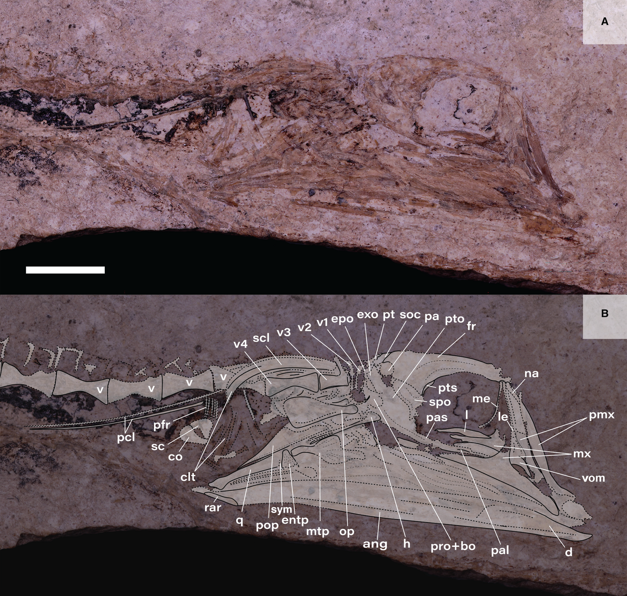
The featured image shows a real Eocene tube-eye fish fossil and a separate interpretative reconstruction. It is geological palaeontology, not human archaeology; the paper itself acknowledges uncertainty around the prootic–basioccipital boundary. The Cove Hazel measurements and bone counts are explicitly fictional classroom examples.
The scientific foundations are ZFIN’s prootic definition, Jessen’s spottail shiner variation study, and the golden grey mullet micro-CT osteology.


Prootic evidence: the claim ladder
| Direct object or finding | Reasonable scientific use | Not established alone |
|---|---|---|
| Prootic contact surface near parasphenoid and basioccipital | Identification of neurocranial region | Unique species without adequate comparison |
| Intact nerve foramen and rim | Taxon-specific anatomical character | Exact hearing acuity or nerve performance |
| Otic recess within skull | Inner-ear enclosure anatomy | Otolith age rings |
| Raw fragmented and complete objects per sediment mass | Description of recovered fossil sample | Fish population or annual catch |
| Bone in settlement sediment | Possible human or non-human source history | Specific meal, trade route or capture date |
1. Identify the prootic before identifying ancient fish behavior
The prootic is commonly a paired endochondral bone contributing to the ventrolateral neurocranium and otic region of many teleost fishes. Its facets, laminae and foramina link the auditory capsule with surrounding skull bones and cranial nerve or vascular passages. A surviving archaeological prootic can be informative where contact surfaces are intact, but one mineralised fragment is not an otolith, a fish age reading or a record of how an ancient animal heard. A species name or a human-use claim requires separate comparison and site context.
Ask students to label the preserved original margins first, and only then compare a possible taxonomic assignment. A little opening might be an actual nerve foramen, an artifact of crushing or a broken boundary. The analyst needs to distinguish each before interpreting fish ecology, fishing technique or a historical meal.
2. Locate the prootic around the ventral otic capsule
In many fishes the prootic lies below the pterotic and sphenotic, behind the pterosphenoid and toward the anterior side of the basioccipital and exoccipital regions. It can form a large wall or floor of the cranial cavity near the internal ear. The exact contacts differ by lineage and developmental stage. A whole skull makes these relationships easy to locate, whereas an isolated fragment may have lost its distinctive articular faces.
Students should map the orbit, ear capsule, prootic, parasphenoid and basioccipital on a source-identified skull, then identify which remaining contacts could confirm an archaeologically detached bone. It is better to record one or two reliable original surfaces than to fill a generic fish-head outline with imagined sutures.
3. Use ZFIN’s definition with its actual taxonomic scope
The Zebrafish Information Network defines the prootic as a paired cartilage-replacement bone contacting the pterosphenoid anteriorly, sphenotic and pterotic dorsally, and posterior cranial bones toward the basioccipital. It also notes foramina associated with trigeminal and facial nerve branches. This is useful standardised anatomical language for a defined reference fish, not a guarantee that every teleost has the same number or arrangement of openings.
Students should copy the definition into a comparison sheet and mark what is directly observable on a modern zebrafish reference. They should then contrast it with a mullet or shiner rather than assume the larval timing or foramen pattern automatically carries over to every archaeological species.
4. Do not confuse the prootic with the pterotic
The pterotic forms a different, often more posterodorsal and lateral region of the otic skull, sometimes carrying part of the lateral-line canal or contact with the posttemporal. The prootic forms a more ventral component and can help enclose neural or vascular channels. Because the two touch, a fractured specimen may appear to contain one irregular plate where two original bones were present. A matched articular reference is critical.
Give students genuine reference views and ask them to identify the original prootic-pterotic interface. If the archaeological contact is damaged, record neurocranial fragment uncertain rather than assign a precise element from position on a loose sorting tray.
5. Distinguish prootic from sphenotic and pterosphenoid
The autosphenotic or sphenotic occupies a more anterolateral area of the braincase near hyomandibular facets, while the pterosphenoid lies around the orbital-braincase wall. Their contacts with the prootic can shape foramina and articular facets, making boundaries difficult to recognise on incomplete material. A perforation shared by two bones should not be coded as wholly enclosed by one unless the relevant suture is visible.
Students should annotate separate original sutures in a modern specimen and mark foramen ownership only after documenting the border. A species comparison based on such openings is valid only when the right bone surfaces survive and the reference covers normal variation.
6. The parasphenoid overlaps the prootic ventrally
In golden grey mullet the parasphenoid extends beneath parts of the prootic, and winglike ascending processes contact the prootic region. A preserved skull may therefore hide part of the prootic from ventral view. If the skull is disarticulated, an exposed facet might look unlike the original complete reference. The relationship is useful for osteology but must be described within its actual fish taxon.
Have students compare the fully articulated mullet neurocranium with drawings in which the parasphenoid is removed. They should record what changed in visibility, without interpreting the newly revealed surface as a new anatomical element.
7. The 2020 golden grey mullet study supplies a real 3D model
Thieme and Moritz combined cleared-and-stained specimens with micro-CT in their 2020 study of *Liza aurata*. Their reconstructions show the prootic as a large ventral cranial component, connected to the parasphenoid, basioccipital, sphenotic and pterotic. This research supports a three-dimensional contact map and highlights that relationships can be missed in ordinary external photographs. It is not a model of all ancient fish skulls.
Students should explain which parts were observed in a prepared specimen and which were reconstructed through segmentation. A CT rendering can be scientifically useful without functioning as a direct image of an excavated fish in the fictional classroom cases.
8. Understand the anterior otic bulla without turning it into an otolith
The golden grey mullet prootic forms part of the anterior otic bulla, while neighbouring skull-base elements contribute elsewhere. An otic bulla is an anatomical enclosure or cavity, whereas a sagitta, asteriscus or lapillus is a mineralised otolith within the ear system. The two should never be counted or aged as though they are identical physical objects. A broken skull cavity may preserve its original outline even after the otolith has been lost.
Students sort photographs of cranial recesses and actual otoliths. They should state which specimen could support a validated growth-ring analysis and why an empty otic cavity itself is not a fish-age measurement.
9. The lapillus recess has specific positional evidence
In the 2020 mullet reconstruction a small hollow dorsal to the otic bulla is identified as associated with the lapillus. This is a useful example of a specific internal cavity linked to a known source fish. Yet the presence, size and exact bony coverage of such recesses differ across taxa. An archaeological depression should not automatically receive this label without correct orientation and comparative evidence.
Ask learners to locate the hollow in the published figure and mark its relationship to the prootic and surrounding otic region. For a damaged specimen, they should retain a broader recess category where the necessary contact is no longer visible.
10. Semicircular canal arches require anatomical context
The prootic can participate in an arch or contact region for the horizontal semicircular canal toward the exoccipital, as documented in golden grey mullet. This identifies where a sensory canal originally passed through the skull, not an individual fish’s hearing or balance acuity. A fossil broken at the canal can show an apparent tunnel whose original path is uncertain. The surrounding otic skeleton is necessary for a responsible reconstruction.
Students should sketch a partial canal and label the verified original surface. They must not infer swimming speed, orientation in water or sound sensitivity from one arch dimension without validated functional and ecological evidence.
11. What a trigeminal-facial chamber means
The trigeminal and facial nerve systems traverse or pass near multiple cranial openings. In some fishes the prootic contributes to a chamber or canal complex that transmits nerves and associated tissues. The precise wall of an opening may be shared by several bones, and surrounding bony struts may vary. One foramen should not be labelled ‘the fish’s hearing hole’ simply because it appears near the ear capsule.
Ask students to distinguish the trigeminal-facial chamber from an otolith recess and the foramen magnum. They should name the source reference that identifies a particular opening, and document whether the original rim is intact before using it in taxonomic comparison.
12. The 2025 spottail shiner study reveals real within-species variation
Erika Jessen studied the spottail shiner *Hudsonius hudsonius* and reported that the anterior opening of the trigeminal-facial chamber could be shared by the prootic and adjacent bones or fully enclosed by the prootic in particular specimens. Some fishes had bony struts near the opening and some differed between left and right sides. This is an unusually valuable caution: an apparently diagnostic number or border of foramina can vary within one fish species.
Have students compare several verified reference individuals before naming one archaeological bone as a different species. A single new-shaped foramen is not sufficient evidence of a new taxon if the published normal range includes such variation.
13. Do not make an asymmetry into a new species
Jessen’s shiner work reported specimens whose corresponding cranial foramina differed between sides. An asymmetrical prootic opening may therefore be normal variation, developmental difference or preparation effect, rather than evidence of pathology or species identity. A fossil preserving only one side cannot show whether the original fish had the same character bilaterally. Classification confidence should reflect that missing comparison.
Give students a paired modern neurocranium with different left and right foramen boundaries. They should describe both sides as observations from one organism and resist the incorrect practice of splitting them into separate taxa.
14. The oculomotor foramen is not a unique prootic signature
The oculomotor nerve opening can be associated with the prootic and pterosphenoid region in particular fishes. In shiner specimens, Jessen documented alternative arrangements in which it was bordered by multiple bones or enclosed differently by the prootic. Its presence and location are anatomically meaningful, but taxonomic inference requires observing the actual contact boundaries and the possible within-species states. A missing edge cannot establish ownership.
Students should draw the foraminal rim in a reference image, annotating each contributing bone where the suture is visible. If one rim segment is eroded, record border uncertain rather than forcing an assignment from a generic diagram.
15. A bony strut may form over a canal in some individuals
The 2025 spottail shiner analysis noted bony struts over the anterior trigeminal region in a subset of specimens. This kind of structural variation can make an opening appear closed or divided in a low-resolution photograph, even when normal nerve pathways remain. It also demonstrates why taxonomic keys based on one reference specimen can overlook ordinary variation. The observer should score strut presence separately from open-passage identity.
Students should record whether a true continuous strut, a broken process or overlap from a neighbouring bone is visible. A provisional foramen character is an honest scientific result when the scan or fossil cannot resolve the distinction.
16. The golden grey mullet anterior myodome is a further context
In the golden grey mullet, the prootics contribute to the roof of an anterior myodome, a space associated with eye-muscle structures within the neurocranial region. Its morphology is described with neighbouring cartilage and parasphenoid relationships. A classroom drawing should not call any ventral skull recess an otic cavity simply because it lies close to the prootic. Anatomical function depends on the actual contact map.
Ask learners to identify a myodome-related region separately from a sensory recess and a nerve canal in the original published figure. These spaces differ despite being inside the head.
17. Prootic contact with the hyomandibular facet
In many fishes, a region near the prootic, sphenotic or pterotic helps form articular facets used by the hyomandibula within the suspensorium. This contact contributes to jaw-supporting anatomy, but an isolated prootic facet does not record the amplitude or speed of jaw movement during life. The functional mechanism includes multiple bones, ligaments and muscles. Taxon-specific references are needed before describing a particular articulation.
Students should label skull-side and hyomandibular-side facets and avoid estimating bite force from one contact surface. A useful anatomical interpretation identifies where the joint belonged without claiming a live feeding event.
18. Historical terminology can be misleading
Nineteenth- and early twentieth-century fish osteology used terms such as petrosal, prootic, opisthotic and alisphenoid in ways that later researchers reassessed. The 1911 salmon chondrocranial drawing and modern reference osteology need a terminology crosswalk rather than silent renaming. Two drawings may refer to corresponding cranial regions with different historical names, while different lineages can further complicate homology.
Have learners keep the original figure labels beside current names and the source taxon. This permits a second specialist to revisit the anatomical interpretation without losing the original evidence.
19. The 2026 Eocene tube-eye fossil limits prootic certainty
Ridolfi and colleagues’ 2026 study of *Parastylephorus anatoides* reconstructed an approximately Eocene fish skull with complex overlap near the prootic and basioccipital. The authors explicitly acknowledged that the exact size and boundary of the prootic were difficult to interpret from the preserved fossil. Their figure groups the region as pro+bo where the anatomy is uncertain. This is a model of honest palaeontological uncertainty, not a weakness to hide.
Students should inspect the actual fossil and interpretative panel separately. A publication’s labelled reconstruction is still an interpretation of physical evidence. They should not convert its approximate boundary into a precise original bone measurement or pretend the Eocene fish belonged to an archaeological human catch.
20. Do not call the Eocene reconstruction human archaeology
The real tube-eye fossil comes from geological strata of the Eocene of Iran, far predating human settlements. It is a genuine physical fossil paired with an expert reconstruction and is useful for teaching which skull bones overlap when flattening obscures boundaries. It does not provide a record of fishing gear, markets or meals. Its geological age is distinct from fish biological age and any fictional classroom deposit chronology.
Ask students to prepare source cards for the fossil, modern reference mullet and an imagined archaeological prootic. The cards should record actual organism, original specimen, time period, method and uncertainty rather than blending these into a single narrative.
21. Foramen preservation has multiple failure modes
A real cranial opening can appear enlarged when its rim breaks; it can appear enclosed by secondary mineral deposits or disappear when a thin lamina collapses. Abrasion, burning, digestion and processing may affect neural canal walls differently from robust articular faces. A diagnosis relying on one incomplete hole is therefore vulnerable to taphonomic bias. Classification should distinguish present, absent on intact surface and unobservable due to damage.
Students should work with comparative photographs showing an original rim and a damaged one, then state which taxonomic claims remain supportable. A small specimen may identify the prootic region without allowing exact foramen counts.
22. Micro-CT helps but does not restore lost anatomy
Micro-computed tomography can show internal skull cavities and help distinguish a true canal from a surface depression when density contrast is adequate. Yet resolution, segmentation thresholds and post-depositional distortion can create apparent false surfaces. The Eocene reconstruction and mullet 3D work illustrate how software supports interpretation rather than replacing anatomical expertise. A single digital view may obscure contacts visible in raw scan slices.
Students should ask for multiple cross sections and original-source metadata before accepting a proposed nerve passage. If the structure is physically missing, an attractive 3D rendering cannot manufacture the original boundary.
23. A prootic is often paired, so sidedness matters
Many fish have distinct left and right prootics. Their individual fragments can be separated during burial and may appear as two unrelated bones to an analyst who lacks reference anatomy. A species diagnosis should record side and internal or external view when possible. Because a single bone can fragment and a fish has two prootics, a raw count of prootic objects is not a count of individuals.
Give learners two secure left prootics and several unsided fragments. They should compute a conservative minimum only when anatomy, taxon and closed context justify it, while keeping physical pieces and refits in a separate table.
24. Geometry of a foramen is not a hearing test
For teaching dimensions, a prootic opening may be described by major and minor axes measured between intact original margins. Such a ratio can be compared within a reference taxon, but it does not quantify hearing sensitivity, balance, fish age or swimming speed. Biological inference requires physiological or experimental validation. Postmortem erosion can alter the shape much more than between-species variation.
Students should draw exact measurement endpoints and list the potential bias caused by a broken rim. Numeric observations should be separated from speculative sensory function.
25. An entirely fictional Cove Hazel geometry exercise
Imagine a toy prootic opening in the fictional Cove Hazel reference fish. Specimen A has major diameter 3.0 mm and minor diameter 1.5 mm, a ratio of 2.00. Specimen B is 4.0 by 2.0 mm, also 2.00. These values are invented and do not belong to the actual mullet, spottail shiner or Eocene fish literature. Matching ratios say nothing by themselves about taxonomic identity or how sensitive a fish’s ears were.
Students should retain both absolute measurements and describe what one ratio does and does not establish. A fish-size model would require modern verified reference measurements and much broader evaluation.
26. Do not use a truncated opening as a full-facet measurement
Suppose an ancient prootic has lost part of the rim, leaving a measurable hole-like depression. The observed length of this damaged remnant is not the original canal’s major axis. A caliper may be precise yet biologically incompatible with the defined reference variable. The same problem arises where adjacent bone sutures were flattened and later mistaken for an opening.
Students should record the preserved dimensions and mark the complete opening as unmeasurable. Withholding an invalid number is better than inventing a missing boundary to fill a worksheet.
27. Sieve and recovery influence cranial-bone abundance
An isolated prootic fragment may be small, brittle or difficult to recognise without osteological comparison. Fine-sieving and deliberate microscope sorting can yield more identifiable material than an older coarse-screened archaeological excavation. Sampling mass, sediment volume, sorting effort and diagnostic thresholds all affect apparent abundance. Raw comparisons across incompatible methods can be mistaken for real biodiversity changes.
Have learners compare two excavation protocols and explain why a taxon’s appearance after improved recovery is not necessarily its first presence in the ancient environment. The reporting standard is to keep the recovery process with the count.
28. A fictional Cove Hazel fragment-count table
In a wholly invented Cove Hazel teaching dataset, the older layer contains twenty-four complete prootics and thirty-six identified fragments from three kilograms of dry sediment. The younger contains eighteen complete and sixty-six fragments from seven kilograms. Total objects are sixty and eighty-four, equivalent to twenty and twelve per kilogram. A higher raw total in the younger layer coexists with a lower concentration under the invented processing rule.
Students should calculate both and label the unit as classified prootic objects per processed dry sediment. The numbers and place name are fictional; they do not measure fish caught or an actual ancient population.
Cove Hazel: fictional foramen measurement
| Imaginary reference | Major axis | Minor axis | Major/minor |
|---|---|---|---|
| A | 3.0 mm | 1.5 mm | 2.00 |
| B | 4.0 mm | 2.0 mm | 2.00 |
| Broken C | Original rim absent | 2.0 mm | Not calculated |
29. Examine completeness separately from recovered totals
In the imaginary older layer complete bones form 24/60, or forty percent, and in the younger 18/84, about 21.4 percent. Complete-object concentrations change from eight to approximately 2.57 per kilogram. The larger contrast for intact material may reflect fragmentation or different preservation conditions rather than fish population change. Both total counts and condition categories should remain visible.
Ask students to propose a taphonomic test for physical abrasion or differential laboratory damage. A precise description of altered completeness is scientifically valuable even if the cause remains unknown.
30. Human settlements can contain naturally introduced prootics
Fish cranial elements may arrive through natural mortality, predator prey, flooding, processing waste or trade in fish products. A prootic found beside pottery is not automatically evidence of cooking, and a marine fish skull bone in an inland settlement may reflect exchange rather than the location where the fish grew. The strongest historical interpretation tests site formation and fish-source ecology separately.
Students should map habitat, death, potential processing, transport, final burial and recovery as distinct stages. They must identify which observation supports each link and avoid making one bone an all-purpose historical witness.
31. Archaeological dates do not come from nerve canals
A prootic’s foramina and articular contacts may inform species identification but generally do not yield the calendar year of deposition. Dating comes from stratigraphy, associated datable evidence and assessment of reworking. A fish bone can be carried into younger sediment after its source organism died. Chronological charts of prootic counts should therefore display age ranges instead of fabricated annual catch sequences.
Students should compare sample depths and a dated layer with broad error bars. They should not assign an exact fishing regulation or climate event to a count shift without independent temporal and cultural support.
Cove Hazel: invented specimen counts
| Invented horizon | Complete bones | Diagnostic fragments | Total objects | Dry sediment | Objects/kg | Complete share |
|---|---|---|---|---|---|---|
| Older | 24 | 36 | 60 | 3 kg | 20 | 40% |
| Younger | 18 | 66 | 84 | 7 kg | 12 | 21.4% |
32. Conservation and attribution complete the evidence chain
Ancient prootics can be fragile and their diagnostic rims irreplaceable. Chemical cleaning, destructive sectioning or casting may alter tiny openings. Appropriate curatorial authority, condition records, photographs and non-destructive methods should precede consumptive analysis. When using published figures, report whether they depict a modern dissection, a digital reconstruction or a geological fossil. Authorship and licence are necessary parts of a reliable public teaching article.
Have learners prepare a specimen and figure audit sheet including museum identifier, method, taxon, geological or archaeological age, source permissions, preserved structures and unresolved characters. A second scholar should be able to assess the same bone without relying on undocumented claims.
33. Write the narrow conclusion that survives peer review
A good prootic report can show a verified neurocranial element with preserved contact faces, document a specific canal boundary where intact, and compare its form against appropriate reference fishes. It cannot independently measure hearing, bite force, annual catch or the behaviour of one fish. The fictitious Cove Hazel rates are a lesson in counting and preservation, not a real ecological trend. The 2025 shiner data also show why within-species variation must be considered.
The gold standard is an auditable anatomical identification with source-linked character states, explicit fragment and sediment denominators, separate cultural hypotheses and an independent test for whatever remains uncertain.
Twenty-six real-world reasoning challenges with fictional specimens
All case specimens and sites in this section are imagined. They are not newly documented archaeological findings.
1. One nerve opening, two species labels
A prootic foramen is bounded differently on the left and right of a known spottail shiner. Students explain why ordinary intraspecific asymmetry can defeat a simple species key.
2. The prootic versus pterotic confusion
A broken lateral otic fragment is labelled pterotic because it is near the skull roof. Students inspect original ventral cranial contacts before assigning bone identity.
3. The sphenotic contact is lost
A fossil’s anterior margin is fractured, eliminating a key contact with the sphenotic. Learners preserve a broad otic-region identity rather than force a species.
4. The parasphenoid wing covers the facet
An articular surface is hidden in a complete skull but visible when bones separate. The student claims a newly evolved structure. Reviewers explain anatomical exposure rather than invented novelty.
5. The lapillus recess mistaken for an otolith
An empty skull cavity is described as a mineralised lapillus suitable for age counting. Students distinguish the otolith itself from its supporting cranial recess.
6. The trigeminal opening in one specimen
A paper documents a foraminal border variation; the learner makes it a universal genus character. The class checks the source’s specimen distribution and confidence.
7. A new bony strut
The 2025 shiner study found bony struts over a nerve opening in a subset of fishes. Learners explain why presence or absence may be variable within a species.
8. The oculomotor border shifts
A shared prootic-pterosphenoid rim is mislabelled entirely prootic. Students inspect the actual suture and record border ownership honestly.
9. A myodome called an auditory canal
A ventral muscular space is catalogued as an inner ear recess. The class uses the original mullet anatomical figure to distinguish distinct cavity functions.
10. The Eocene pro+bo label
A reconstruction combines prootic and basioccipital near a damaged contact. Students explain why the authors did not pretend to know a complete boundary.
11. Ancient fossil presented as human fishing
The Eocene tube-eye fish skull is captioned as a settlement catch. Students correct the geology and separate source anatomy from invented field data.
12. The blue 3D rendering trap
The coloured prootic in a published CT reconstruction is treated as a photograph of an isolated whole bone. Students distinguish segmentation from raw physical specimen images.
13. A low-resolution hole
An imaging artifact appears as a new nerve foramen. Learners request original sections and preserved rim evidence before diagnosing a taxon.
14. A damaged margin appears smooth
An abraded articular facet is called a biological absence of a process. Students separate unknown preservation state from observed true anatomy.
15. Two paired prootics from one fish
Right and left elements are counted as separate animals. Learners map paired skeletal source before computing any minimum individual number.
16. One bone becomes several fragments
A prootic fractures into four diagnostic pieces during sorting. Students document raw physical count and possible refits without multiplying fish.
17. The equal-ratio illusion
Two imaginary prootic openings yield major/minor ratio two at different absolute sizes. Students explain why the ratio is not a fish-length or hearing-performance measure.
18. The missing canal rim
A hole-like fragment retains only one original measurement endpoint. Learners preserve observable dimensions and withhold the full shape ratio.
19. The raw count reversal
Cove Hazel has 60 objects from 3 kg older and 84 from 7 kg younger. Students calculate twenty versus twelve per kilogram, refusing a population story.
20. Complete share changes
The fictional intact fraction falls from 40 to 21.4 percent. Learners investigate transport, chemistry and specimen handling before ecological attribution.
21. Fine sieve discovers new small bones
A new excavation retrieves tiny prootic pieces absent in coarse-screened older reports. The class tests recovery bias before declaring fish arrival.
22. A marine fish at an inland midden
A secure marine prootic occurs in a town dump. Students consider fish trading and food transport instead of inventing a local ancient sea.
23. Predator accumulation near pottery
Fish cranial fragments have possible digestive modification. Learners contrast predator feeding with human preparation using independent sediment context.
24. One historical fishing year invented
A prootic count changes across broad archaeological age intervals. Students remove an unsupported exact-date causal claim and plot proper chronology.
25. A wrong skeletal model input
A cleithrum body-size regression is given a prootic hole width. Reviewers reject the anatomical mismatch, not merely the numeric precision.
26. The final audit
One damaged prootic supposedly proves species, ear acuity, prey, fishery abundance and capture technique. Students keep original observations and identify separate tests for every added inference.
Three-session osteology and archaeology teaching sequence
- Session 1: map prootic, prootic foramina, parasphenoid, sphenotic, pterotic and basioccipital on labelled real-source skull views, separating skull recesses from otoliths.
- Session 2: compare spottail shiner within-species foraminal variation and modern mullet 3D otic architecture with the uncertain Eocene pro+bo interpretation.
- Session 3: compute fictional Cove Hazel counts and geometry, assess screened sediment, fragments and original elements, and test cultural versus natural source hypotheses.
- Submission: scaled source image, diagnostic character table with confidence, original data and mass denominators, alternative depositional pathways and one targeted next study.
Gold Standard scientific assessment rubric
| Scientific task | Evidence that meets the standard | Overclaim to avoid |
|---|---|---|
| Bone identification | Correct prootic and neighbouring skull contacts | All irregular holes called prootic nerve foramina |
| Biological comparison | Within-species and bilateral variation considered | One unusual opening identified as new fish species |
| Sensory interpretation | Otic enclosure distinguished from otolith and function | Hearing capacity inferred from skull cavity size |
| Quantitative reporting | Original rims, bones, fragments, sample mass and limits recorded | Fossil pieces treated as independent fish |
| Archaeological reasoning | Predator, trade, natural transport, human discard and chronology assessed | Bone near pottery automatically called human catch |
| Research image | Actual fossil versus reconstruction and licence explained | Eocene specimen passed off as human archaeology |
Frequently asked questions
What is a fish prootic bone?
A typically paired bone of the ventrolateral braincase and inner-ear region, associated with cranial foramina and other skull bones.
Is the prootic an otolith?
No. Prootic is part of the skull; otoliths are separate mineralised ear structures.
Can a prootic identify fish species?
Sometimes through preserved diagnostic contacts or openings, but within-species variation must be considered.
What is the trigeminal-facial chamber?
A cranial passage associated with nerve and related structures that may involve the prootic and neighbouring bones in different arrangements.
Do all fishes have identical foramina?
No. The 2025 spottail shiner study even documents variation among specimens and between sides of individuals.
Can a prootic measure hearing sensitivity?
Not directly. One fossil cavity cannot quantify hearing or balance performance without physiological validation.
What does pro+bo mean on the 2026 fossil figure?
An interpreted region involving prootic and basioccipital whose exact boundaries are difficult to determine on the fossil.
Is the featured Eocene fossil human archaeology?
No. It is a geological fossil and an associated reconstruction, not a historic fishing specimen.
Are Cove Hazel’s rates actual measurements?
No. Cove Hazel and its specimen data are invented for the classroom.
Does one fragment represent one fish?
No. Paired bones can fracture, and counts of recovered skeletal pieces differ from original animals.
Scientific sources, original figure and connected eduKate research
- ZFIN prootic anatomy ontology ZFA:0000575
- Jessen (2025), spottail shiner prootic, trigeminal-facial openings and variable foramina
- Thieme & Moritz (2020), golden grey mullet inner ear and 3D neurocranium
- Ridolfi et al. (2026), actual Eocene skull and uncertain prootic+basioccipital region
- Original fossil and interpretation reference image, CC BY 4.0
- Rojo (2013), Nova Scotia brown bullhead skull-base osteology
- Ossification of Inpaichthys kerri fish cranial regions
- Development of zebrafish craniofacial bones in 3D
- The Gold Standard of Teaching Archaeological Fish Basioccipital Analysis
- The Gold Standard of Teaching Archaeological Fish Otolith Analysis
- The Gold Standard of Teaching Archaeological Fish Parasphenoid Analysis
- The Gold Standard of Teaching Archaeological Fish Hyomandibula Analysis
- The Gold Standard of Palaeoecology
- The Gold Standard master hub
The gold standard of prootic analysis is a learner who identifies the proper otic skull bone, records which cranial nerve openings actually survive, understands how normal within-species variation affects diagnostic claims and separates fossil counts from fish populations. Geological reconstructions, living-fish references and archaeological contexts can inform each other without becoming interchangeable evidence.
Foramen-code sensitivity clinic
Suppose a damaged opening is categorised first as absent, then properly changed to indeterminate after inspecting the original photograph. Students explain how a species diagnosis and a numerical character matrix may change even though the physical specimen has not. They should preserve both recorded decisions and the source of the correction.
CT representation and reality
A segmented 3D model colours the prootic dark blue, giving the appearance of a complete plate. Students return to original source slices and recognise that some borders are algorithmic or interpretative. A digitally sharp boundary is not always a physically preserved suture.
Museum custody and two-age problem
An Eocene fossil has a geological date, but its individual fish age-at-death remains unknown. An archaeological bone has a later sediment context that may also differ from its death. Students distinguish source chronology, individual growth and final deposition to prevent an exact-year claim from a skull feature.
Inter-reader agreement
Two analysts identify a partially preserved nerve passage differently. Students should record which rim they each saw, compare a documented modern reference and reach a confidence-qualified diagnosis. Agreement alone is not evidence of correctness when both depend on one inappropriate comparison.