
The retroarticular is a small element associated with the posterior lower jaw in many ray-finned fishes. Its shape, contacts and degree of separateness vary among fish lineages, making it a useful but sometimes difficult object of archaeological identification. The bone may help reconstruct original jaw anatomy but cannot by itself reveal how many fish lived at a site, what one fish ate or how it was caught. Identification, taphonomy and cultural interpretation require separate evidence.
This complete reference develops skeletal location, confusing neighbouring bones, taxon-dependent development, possible ligament associations, archaeological recovery and a transparent fictional River Larch calculation. The scientific figure compares a true Eocene tube-eye fish and a modern living relative; it is not material excavated at River Larch or associated with human occupation. The chapter index and evidence table help readers reach the answer appropriate to the observation.
The most relevant sources are the 2026 paired stylephorid skull study and the review of fish jaw development. Anatomical labels and archaeological specimen-count methods should be treated as separate kinds of evidence.


What the evidence means at a glance
| Observed object | Possible conclusion | Not established by that object alone |
|---|---|---|
| Small posterior mandibular ossification | Broad anatomical or retroarticular diagnosis | One named species or one fish |
| Apparent articular or ligament-contact facet | A source-appropriate contact hypothesis | Measured feeding speed |
| Several complete or fragmentary pieces | Inventory with explicit unit and context | Original number of animals |
| Two measured original landmarks | A descriptive shape ratio | Fish body length without modern calibration |
| Bone from a human-associated sediment layer | A candidate human activity explanation | One specific meal or date |
1. Begin with the tiny posterior bone, not a fishing story
The retroarticular is a small skeletal component associated with the ventral or posterior end of the lower jaw in many ray-finned fishes. Its morphology, separateness and contacts vary between lineages, so it is not a universal one-shape bone. In archaeological contexts it may survive attached to the jaw or as a loose fragment. Identifying it can improve understanding of skeletal anatomy, but one little bone is not one animal, a tooth, a measured bite force or independent proof of a human catch.
Begin with an observation ladder. A small element is present at the posterior jaw; a specialist identifies it as retroarticular; its source taxon is inferred; ancient people are said to have caught that fish. Each rung requires new evidence. Learners should annotate original anatomical position and diagnostic comparison first, and treat the proposed human activity as a separate question about how the bone reached the excavated layer.
2. Locate retroarticular behind the dentary
In many teleosts the dentary forms much of the anterior lower jaw while the anguloarticular contributes to the posterior primary joint. The retroarticular occupies a smaller region near the ventroposterior jaw, where its contact with adjoining elements can be taxonomically informative. The position is not exactly identical in all fish groups. Removing surrounding bones can make a detached retroarticular look like an indeterminate pebble or process, particularly when the original margins are abraded.
Students should outline dentary, anguloarticular and retroarticular on a properly labelled jaw before examining the isolated object. They record which neighbour would have contacted each preserved facet. If the original association cannot be reconstructed, an uncertain posterior-mandibular identification is stronger than guessing a small species-specific element from shape alone.
3. Learn the difference from anguloarticular
The anguloarticular generally includes the main lower-jaw articulation with the quadrate in many teleosts; the retroarticular is a smaller neighbouring element and should not be counted as a second copy of that joint. Published fish anatomical descriptions distinguish these terms, but historical naming and fusion can vary. A composite fragment may show several elements joined, and one bone can separate at a pre-existing suture during burial.
Provide students with an intact lower jaw and a partly separated version. They must identify whether the articular socket belongs to the anguloarticular and where a smaller retroarticular originally sat. This teaches a useful distinction: physical proximity does not make two skeletal categories interchangeable. A correct joint comparison also prevents double counting original fish.
4. Distinguish the quadrate as the opposing joint bone
The quadrate forms the opposing upper component of the jaw joint in many bony fishes, contacting the anguloarticular. It is not the retroarticular, even though several elements cluster near the back of the mandible. A detached quadrate process can resemble a small jawbone fragment if it has lost its distinctive articular condyle. Source anatomy is essential for separating these parts from one another.
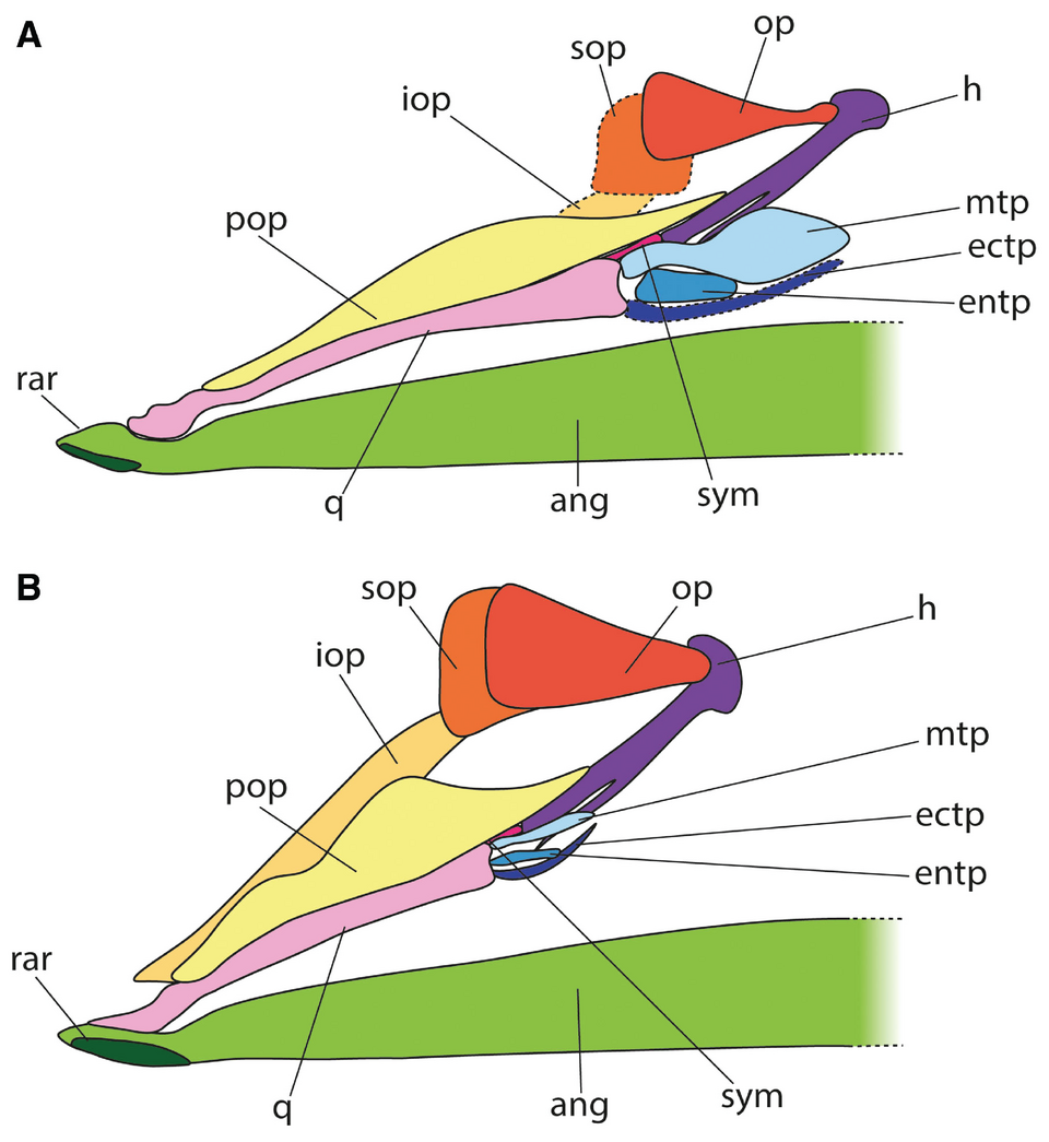
Students should identify q, ang and rar in the featured comparative figure and explain that each abbreviation names a distinct anatomical component. They then hypothesise which features remain after a fragment separates from its neighbours. The task rewards correct element identification before species naming or functional claims.
5. The featured stylephorid comparison is not a human excavation
The scientific figure reproduced here compares posterior skull anatomy in an extinct Eocene tube-eye fish, Parastylephorus anatoides, and the living Stylephorus chordatus. It explicitly marks the retroarticular as rar. The 2026 research is valuable for comparative morphology and understanding fish evolutionary history, but the ancient Eocene specimen is geological palaeontology, tens of millions of years older than any human-associated archaeological fishery being discussed in the classroom.
Ask a learner to read the A and B panel labels, annotate retroarticular and anguloarticular and identify which image is an interpretive reconstruction. A correct figure caption must retain the authors, fossil-versus-modern contrast and Creative Commons licence. It must never describe this plate as the newly excavated contents of the fictional River Larch case.
6. Compare the small element with the whole jaw
A loose small bone carries much less contextual information than the same element observed within a complete skull. Contact surfaces that seem ordinary when isolated may become distinctive through their position relative to the anguloarticular and the interopercular apparatus. Some taxa show altered fusion and spacing of these parts. Consequently, one photograph taken from a single angle may not be a sufficient identification reference.
Prepare paired cards showing a known jaw before disarticulation and its retroarticular after removal. Students identify the contact lost from the isolated photograph and document side, scale and orientation. A useful catalogue may reach element level while the source species remains unknown.
7. Historical names can obscure modern homology
Older ichthyological texts may apply angular, articular or retroarticular to regions and bones using conventions that differ from current taxon-specific terminology. An anatomical name may be based on adult position, while modern developmental research resolves a separate origin or fusion history. Researchers should record exactly how a source defines the element before reconciling old and new catalogues. Replacing every historic label mechanically risks merging dissimilar bones.
Give students two historical descriptions of one fish jaw that use different terms. They should build a terminology crosswalk from original illustrations, modern definitions and actual observed contacts, retaining uncertainty when homology cannot be established. Taxonomy is strengthened by making language traceable, not by hiding a disputed identity.
8. Consider sesamoid and ligament-related interpretations carefully
Small skeletal elements near tendons or ligaments may be described as sesamoid-like in some fish lineages, and their relationship to the interopercular apparatus can be discussed in comparative osteology. Yet the exact developmental identity of a retroarticular is not universal. A tiny mineralised object found close to a jaw should not automatically be named a retroarticular or assigned a tendon function without anatomical and developmental reference.
Students can compare a documented retroarticular with an unidentified small ossicle from an unrelated fish. They should use original position, characteristic surfaces and taxon-specific literature, explicitly distinguishing observations from competing developmental hypotheses. A descriptive category such as small posterior jaw ossification may be more scientifically accurate for damaged material.
9. Understand interopercular linkage without inventing motion
In many fishes soft-tissue connections help coordinate the lower jaw, hyoid region and opercular elements. The interopercle is a distinct dermal gill-cover element, not another name for the retroarticular. Comparative skeletal sources sometimes illustrate connections in the posterior mandibular region, but intact ligaments rarely survive archaeological deposition. Their specific attachment geometry cannot always be inferred from a bare bone.
Ask the group to map potential soft-tissue pathways on an appropriate modern reference, marking observed attachment surfaces separately from inferred ligaments. They should avoid claims that a detached fossil records how fast its owner opened the mouth. Living biomechanics requires far more than one posterior ossicle.
10. Learn how jaw opening differs among fish groups
Different fish groups use different coordinated mechanisms to expand the mouth and move opercular tissues. A retroarticular position may constrain one aspect of the lower-jaw linkage, but its presence is not a universal predictor of gape, swimming speed or suction force. Skeletal mechanics depends on the quadrate joint, dentary and anguloarticular geometry, other cranial elements, muscles and ligaments, as well as the animal’s behaviour.
Give students two identified living-fish references with different jaw mechanics. They must state which anatomical comparisons are reasonable and which dynamic variables were never measured in the fossil. A robust report may describe likely original attachment instead of manufacturing a bite-force number.
11. Deep-time tube-eye fossils are useful anatomical contrasts
The Eocene Parastylephorus and living Stylephorus are specialised forms with very distinctive skull geometry. The 2026 comparative plate shows the position of the retroarticular relative to an elongated lower jaw and other suspensorial bones. This comparison can teach that anatomical relationships change in evolution. It should not lead to the assertion that all ancient fishes possessed the same posterior jaw shape or feeding mode.
Learners should write a short comparative observation: which bones occupy similar topological positions in the two source lineages? Then they must separately state what would be needed to interpret the diet or mechanics of an unrelated archaeological fish. Evolutionary comparison informs hypotheses without replacing source-specific evidence.
12. Differentiate biological age from geological age
A fossil described as approximately 40 million years old refers to the geological age of its source strata, while a fish’s biological age refers to the years it lived. Neither is determined from the visible presence of a retroarticular. Archaeological chronology depends on dates and context, and a small jaw bone usually carries no direct calendar timestamp. Confusing these quantities can turn a legitimate evolutionary source into a fictional human historical event.
Have students construct a three-column sheet for geological source age, individual fish age and date of archaeological deposition. They must not copy a time from one column to another without a validated link. The exercise is particularly important when illustrated sources span modern anatomy, Eocene fossils and human settlements.
13. Developmental stages can change bone separateness
In fish ontogeny, jaw components ossify, contact or fuse at species-dependent stages. Comparative studies of clupeocephalan anatomy and newer characiform growth series demonstrate that bones around the angular-articular region do not all develop in identical ways across fishes. A posterior jaw ossicle from a juvenile may therefore appear different from the adult reference. Distinguishing development from taxonomy requires a series of known-age or known-size modern specimens.
Students compare adult and juvenile reference jaws and record whether the retroarticular is discrete, contacting or obscured. A missing separate bone in one image need not indicate that its source species lacked the anatomical region; it could reflect growth, fusion or incomplete preparation.
14. Avoid mistaking breakage for natural separation
A posterior jaw can fracture along a true contact or across a continuous bone. Under magnification, a natural articular surface may show a different contour and texture from a fresh crack, but abrasion and burial chemistry can obscure that distinction. A tiny fragment near the anguloarticular should not receive a separate catalogue entry as a retroarticular without checking whether it belongs to a broken process of another element.
Ask students to photograph the contact area with a scale and assign a confidence class. When refitting is possible, document the original arrangement before finalising the NISP tally. An uncertain fragment is still evidence and need not be given a false precise identity to remain in the archive.
15. Bone side and orientation matter
Left and right lower-jaw elements are often paired and can mirror one another. A fragment viewed medially instead of laterally may seem to have a different curvature or process direction, while a compressed specimen can lose recognisable orientation entirely. Historical plates sometimes use the right jaw and modern atlases the left. Comparisons require explicit side, view and source taxon so readers know which features are genuinely corresponding.
Students should take two photographs of a labelled modern reference and mark dorsal, ventral, anterior and posterior. If side remains indeterminate in the archaeological piece, they should keep that limitation explicit rather than building a minimum-fish count on an invented sidedness.
16. Use a broader element category where needed
Some archaeological fish bones are too small or damaged for a secure retroarticular identification but clearly belong to the posterior lower jaw. That broader identification is useful for establishing skeletal representation and for retaining potentially diagnostic fragments for future comparison. Forcing species precision can bias an assemblage more than reporting uncertainty does. The best taxonomic resolution is the finest one that the actual preserved characters support.
Provide three sample cards with secure, probable and indeterminate identifications. Students should justify each using named original contacts, not how well an object visually resembles a famous specimen. A broad jaw-region category is an accepted scientific finding when the decisive character is missing.
17. Distinguish morphology from taxonomic exclusivity
One particular shape may occur in more than one fish species through shared ancestry, similar mechanics or ordinary within-species variation. A study that depicts one unusual retroarticular does not establish that the bone is globally unique. Comparative identification requires multiple candidate taxa and preferably specimens spanning ontogenetic and geographic variation. A similarity score from one photograph cannot eliminate taxa absent from the reference collection.
Give learners a hypothetical fish from a river outside the reference study’s region. They should identify which local alternatives need checking and why a family-level diagnosis may be stronger than choosing one pictured extinct stylephorid. Source coverage is a real limit on every classification.
18. Count complete retroarticulars separately from fragments
A fish may have two retroarticular regions and each original bone may break into multiple pieces. Thus the number of physical fragments differs from the number of original elements and from the number of fish. A catalogue should separate complete elements, diagnostic fragments, uncertain pieces and possible refits. The raw observation table should remain available even if researchers later compute a conservative estimate of original elements.
Have learners build a mock tray with six complete bones, four fragments potentially refitting to two more, and several unidentified pieces. They must state what is known rather than divide the total by an arbitrary number of bones per fish.
19. Learn when a minimum number of individuals is defensible
Minimum number of individuals is a conservative estimate based on secure anatomical repetition and contextual grouping. Several independently identified right retroarticulars of one taxon within a closed deposit may help constrain a minimum if each is truly distinct and not a fragment of another. An accumulation spanning centuries or an unsided assemblage complicates that logic. MNI is not simply NISP divided by two.
Ask students to contrast three confirmed right elements with fifteen fragments whose original sides are unknown. They should record the former as a potential lower bound only under stated conditions, and retain the latter as specimens rather than additional fish. A transparent definition of context matters as much as the arithmetic.
20. Sieve choice controls recovery of tiny elements
Retroarticular fragments may be small and inconspicuous, making them vulnerable to loss during coarse sieving or inadequate laboratory sorting. Two archaeological deposits recovered with different mesh sizes can appear to have radically different frequencies of posterior jaw bones despite similar original deposition. Sample dry mass or volume, screen mesh, sorting effort and identification protocols must accompany any comparative rate.
Present two fictional site logs, one finely wet-sieved and one hand-collected. Students should state which comparisons remain valid and propose a harmonised reanalysis if suitable archived sediment exists. A better recovery protocol is not proof of greater ancient fish abundance.
21. Describe taphonomic abrasion and chemical change
Surface rounding, mineral dissolution, burning, pressure and predator digestion can alter an anguloarticular-retroarticular region. Diagnostic joint surfaces may survive differently from small tips, creating selective representation. An isolated smooth ossicle might be an originally separate bone or a process made unrecognisable by weathering. The analysis needs condition categories and appropriate comparisons to avoid reading burial damage as a biological trait.
Students classify intact margins, fracture surfaces, abrasion and surface loss on an invented set. They should determine whether the resulting taxonomic pattern changes when damaged specimens are withheld from species-level classification. Taphonomy is part of the measurement process, not an optional caveat.
22. Build a transparent geometry exercise
A dimensionless ratio may be useful for describing the shape of a fully preserved bone, but only when two original landmarks are well defined. Imagine fictional Reference A has a ventral projection length of 1.2 mm and an overall width of 3.0 mm, giving a ratio of 0.40. Fictional Reference B has 1.5 mm and 3.75 mm, also 0.40. Identical ratios do not establish identical species, fish size or jaw mechanics.
Students should annotate both measured lines and explain why scaling both dimensions can preserve the ratio. They must not convert 0.40 into a body length or a fish age; no such relationship has been calibrated for this teaching case. These are explicitly invented specimens, not measurements from the Eocene research image.
23. Withhold a ratio when the original tip is missing
A broken posterior tip can leave a shorter preserved projection than the original full dimension. Using the remnant in a ratio gives a precise-looking but incompatible measurement. Estimating the missing end from a convenient illustration is not equivalent to measuring it. The original reference should specify what to do with damaged specimens rather than assume every fossil is complete.
Provide a specimen where overall width survives but the projection’s endpoint is lost. Students retain width as an observed quantity and mark the ratio unavailable. This is a useful result because the specimen may remain identifiable while being unfit for a particular quantitative analysis.
24. The imaginary River Larch sediment comparison
Consider an explicitly fictional River Larch archaeological sample with forty complete retroarticular objects and twenty diagnostic fragments recovered from two kilograms of processed sediment. A later imagined layer yields thirty complete elements and fifty fragments from five kilograms. Total classifiable objects are sixty and eighty, giving observed concentrations of thirty and sixteen objects per kilogram. The larger raw count in the later layer does not imply a larger original fish population.
Students should calculate both rates and state the measurement unit. The deposit name and all numbers are invented; this is not a case report from an actual river or human fishery. Raw specimens, complete elements and biological individuals must not be treated as interchangeable.
River Larch: invented original-landmark comparison
| Imaginary specimen | Posterior projection length | Original bone width | Ratio |
|---|---|---|---|
| A | 1.20 mm | 3.00 mm | 0.40 |
| B | 1.50 mm | 3.75 mm | 0.40 |
| Broken C | Endpoint lost | 3.00 mm | Not calculated |
25. River Larch completeness changes in a different direction
In the fictional older layer, forty of sixty objects are complete, or about 66.7 percent. In the younger layer thirty of eighty are complete, or 37.5 percent. Complete-object concentrations are twenty and six per kilogram respectively, a sharper change than the overall count rates. This suggests that preservation or breakage should be evaluated alongside any ecological hypothesis. The ratios cannot determine which process caused the observed difference.
Ask learners to graph total objects per kilogram and complete objects per kilogram separately. They should then propose an independent test involving sediment energy, preparation records or fragmentation morphology. A mathematically correct trend is not proof of its cause.
26. Source of fish material is distinct from the findspot
Posterior jaw remains can reach an archaeological layer after natural fish mortality, predator feeding, human processing, trade in fish products or water transport. A marine taxon’s fragment in an inland settlement may indicate trade rather than a local marine habitat. Likewise, river floods can carry bones into an intermittently dry ditch. Anatomical identity cannot independently identify where the original fish lived and died.
Make students map the possible stages from source habitat through death, processing, transport and final deposition. They should identify which independent artefacts or sediment features would distinguish local catch, imported products and natural movement. The most useful historical argument tests those alternatives.
27. Do not infer diet or a meal from jaw support alone
An isolated retroarticular does not preserve tooth wear, stomach contents or feeding events, and its possible association with jaw-opening ligaments does not reveal what a fish ate. A bone found near a hearth may be part of fish preparation waste, but burning can occur after discard and predators can create similar accumulations. Human-use claims require anatomical assemblage patterns, context, marks and chronology.
Give the class a hypothetical small jaw fragment in a drainage lens near pottery. Students should describe a natural pathway and a human pathway, naming a discriminating observation for each. Proximity is a clue, not a sufficient causal explanation.
River Larch: fictional archaeological observations
| Invented layer | Complete objects | Fragmentary objects | Total pieces | Dry sediment | Objects/kg | Complete share |
|---|---|---|---|---|---|---|
| Older | 40 | 20 | 60 | 2 kg | 30 | 66.7% |
| Younger | 30 | 50 | 80 | 5 kg | 16 | 37.5% |
28. Do not assign a precise historic date from bone morphology
The geological age of an Eocene reference fossil is unrelated to the date of an archaeological jaw fragment that happens to resemble it. The archaeological specimen’s time context comes from stratigraphy, suitable scientific dating and an assessment of later disturbance. A fragment can be redeposited in younger sediment long after the source organism died, and some occupation layers integrate many years.
Students should label the physical sample depth, chronological uncertainty and possible reworking. They should not connect one object peak to a named historical flood or fishing law unless independent evidence and time resolution warrant it.
29. The evolution of the posterior jaw requires careful homology
Fish developmental and evolutionary literature describes changing angular, articular and retroarticular relationships. The fact that two bones share a historical name does not mean they have exactly the same development or role in every lineage. Likewise, the mammalian middle-ear evolution literature concerns broad transformations of ancestral jaw elements and cannot be read as the modern function of a recently living fish’s retroarticular.
Ask students to distinguish a deep-time evolutionary comparison from a taxonomic study of a freshwater fish in archaeology. They should record organism, lineage, geological period and the actual comparison supported by each source. This guards against spectacular but inaccurate statements about one tiny bone.
30. Protect original context and conservation history
Rare fish cranial elements may belong to protected archaeological collections. Destructive preparation should follow specimen-owner permission and be justified by a question that cannot be answered non-destructively. Multi-view photography, original reference illustrations and careful catalogue reconciliation may produce more value than consuming one tiny fragment. Conservants or earlier handling may also affect surface interpretation.
Students prepare a custody record that includes site, sediment unit, original catalogue labels, bone condition, orientation, reference images, proposed tests and retained material. The goal is an evidence trail a second researcher can audit without relying on the first analyst’s memory.
31. Publish a conclusion bounded by the evidence
The imaginary River Larch exercise produces a correct contrast in recovered object rates and completeness under deliberately simplified rules. It does not establish fish population decline, a precise species replacement, jaw-opening speed or a particular ancient meal. A genuinely identified retroarticular can add value to a faunal inventory, especially where joint anatomy and modern reference material survive. Taphonomic controls, secure dating and independent archaeological indicators are then needed for more detailed histories.
A gold-standard report states the original observation, identification confidence, count units, preservation and source alternatives. It proposes a specific follow-up test rather than treating unknowns as minor inconveniences. An accurate modest result is better than a polished unsupported reconstruction.
Twenty-six original archaeological decision cases
Each case is invented for teaching. Its findings are not additional published archaeological discoveries; the purpose is to practise identification and evidential judgment.
1. The tiny jawbone mislabelled quadrate
A curved posterior fragment is placed in the quadrate category because it lies near the jaw joint. Students identify which side of the joint it would belong to and compare q, ang and rar in a genuine reference plate. They should retain uncertainty if the original articular contacts are lost.
2. Two labels for one original object
A historical catalogue calls a fragment angular while a new analyst calls it retroarticular. Students examine the source taxon and original naming convention before deciding whether it is one bone or two different structures. Both labels should remain traceable in the archive.
3. The sesamoid assumption
A small ossicle lies near the lower jaw and is immediately called a ligament sesamoid. Learners identify what developmental or anatomical evidence a taxon-specific reference would need before establishing that origin.
4. A juvenile bone appears absent
A juvenile lower jaw lacks a conspicuous discrete retroarticular in one preparation. Students distinguish delayed ossification, fusion, poor imaging and true absence rather than naming a new species from one stage.
5. The fossil stylephorid image is described as archaeology
A 2026 Eocene comparative figure becomes an illustration of a Mesolithic fishing settlement. Students correct the geological timescale, pictured lineages, figure labels and licence. Proper attribution is part of scientific evidence.
6. The jaw-opening speed fiction
An author computes mouth-opening velocity from the length of one retroarticular. Students identify the missing muscles, ligaments, full joint and live motion data, then replace the number with a bounded anatomical statement.
7. The mirrored lower jaw
Left and right fragments look unlike each other in differently oriented photographs. The class aligns source views and records sidedness before declaring separate species.
8. One spicule-like bone actually a process
An isolated mineralised shard is catalogued as a retroarticular, but refitting shows it broke from an anguloarticular process. Students preserve the physical-piece observations and correct the original-element diagnosis.
9. The 0.40 shape-ratio temptation
The invented ventral process length of 1.2 mm divided by 3.0 mm overall width is 0.40. Students explain why this cannot become total fish length or species identity absent calibration.
10. Equal ratios do not mean equal fish size
A second imaginary bone has both dimensions 25 percent larger and also yields 0.40. Learners distinguish geometric similarity from absolute size, taxonomy and growth stage.
11. A broken projection endpoint
A fragment preserves total width but not the original ventral projection’s tip. Students keep the original measurable width and withhold the defined ratio rather than guessing the lost material.
12. NISP versus original elements
One retroarticular is broken into four pieces during sorting. Learners count physical objects, record their possible refit and explain why four pieces do not prove four separate fishes.
13. Repeated right elements
Three securely identified right retroarticulars may constrain a minimum in a closed single-taxon context, but several unsided shards cannot simply increase the minimum. Students document context and independence.
14. The raw count reversal
The fictional older sediment gives sixty objects from two kilograms and younger gives eighty from five. Students calculate thirty versus sixteen objects per kilogram and state why these are not yearly fish catch numbers.
15. Preservation differs between layers
Complete fractions fall from about 66.7 to 37.5 percent in the toy dataset. The class investigates transport abrasion, mineral loss and preparation damage before choosing an ecological explanation.
16. The mesh-size mismatch
Small posterior jaw fragments are retained in one fine-sieved excavation but largely missed by a coarser method. Students diagnose sampling bias before interpreting a faunal disappearance.
17. A marine fish found inland
A secure marine retroarticular is recovered from a town dump. Learners map trade, stored fish products, transported debris and actual original habitat rather than inventing a former coastline.
18. A predator-derived assemblage
Posterior fish-jaw elements cluster in material showing digestive modification. Students compare predator and human discard routes before calling the deposit household food waste.
19. The burned piece near a hearth
A retroarticular is darkened near an archaeological fire feature. The class considers burning during cooking, secondary rubbish burning and natural exposure, with additional context needed for attribution.
20. The exact-year fallacy
A count change spans broad dated sediment intervals but is reported as one historic fishing law’s immediate effect. Students restore chronological uncertainty and distinguish correlation from causation.
21. A reference collection misses the local fish
An African river fragment is identified to a South American stylephorid solely because that is the pictured comparison. Learners request local taxonomic references and keep a broader category until alternatives can be excluded.
22. Destructive sampling without a gain
A rare retroarticular is proposed for sectioning even though its lost original contacts cannot be reconstructed. Students weigh permission, specimen value and non-destructive study before deciding.
23. The body-length model from the wrong bone
A regression for dentary height is supplied with retroarticular width. Learners reject the incompatible anatomical variable even though both are in millimetres.
24. The reproducibility handover
A colleague receives only a colourful fragment count chart with no original images, mass or element coding. Students write a complete metadata request before attempting independent verification.
25. An unsupported evolutionary slogan
A fossil retroarticular is described as a direct human ear bone. Learners differentiate the fish lower jaw’s actual identity from broad mammalian and tetrapod transformations, stating lineage and time.
26. The final River Larch manuscript audit
Students retain the two invented observation rates but remove unsupported claims about the source population, exact fishery method and history. They propose one targeted taphonomic test and one anatomical-reference comparison.
Three-session learning sequence
- Session 1: identify retroarticular, anguloarticular, dentary and quadrate using credible comparative skeletal sources, including Eocene-versus-modern anatomical drawings with provenance.
- Session 2: examine naming conventions, development, possible ligament connections, bone sidedness and taphonomy; solve River Larch’s fictional landmark and count comparisons.
- Session 3: test archaeological screening, NISP and cautious MNI, human-versus-predator sources, imported fish, chronology, licensing and collection stewardship.
- Submission: labelled scale references, specimen condition and confidence, raw fragments plus denominators, two source hypotheses, one feasible independent test and a defensible conclusion.
Gold Standard assessment rubric
| Question | Scientific quality requirement | Shortcut to reject |
|---|---|---|
| What element is this? | Original jaw contacts, side and a matching modern taxon reference | Any tiny curved fragment declared retroarticular |
| How did it develop? | Taxon-dependent source and terminology limitations stated | One growth state used as global species rule |
| Can morphology predict function? | Full-jaw modern biomechanics and appropriate anatomy | Mouth-opening speed invented from one tiny bone |
| What is counted? | Complete elements, fragments, refits, sampling mass and condition distinguished | Every loose piece counted as a fish |
| Is a cultural claim justified? | Independent processing, transport, predator and dating evidence | Bone near artefacts automatically called dinner |
| Is the image accurate? | Fossil-versus-modern original figure, licence and research source retained | Eocene palaeontology falsely labelled human archaeology |
Frequently asked questions
What is a retroarticular in fish anatomy?
A small element in the posterior or ventral lower-jaw region of many ray-finned fishes, whose development and contacts can vary with lineage.
Is it the same bone as the anguloarticular?
No. They are typically described as different components, although historical terminology, fusion and preservation can complicate their distinction.
Where is the retroarticular in the featured scientific figure?
It is labelled rar at the posterior or ventral end of the lower jaw in the two stylephorid comparative reconstructions.
Is that Eocene fish a human archaeological find?
No. The extinct Parastylephorus figure is geological palaeontology, not evidence of a human fishing settlement.
Can the bone identify one fish species?
Sometimes only if genuine diagnostic features survive and the comparative reference adequately covers plausible taxa.
Does one retroarticular mean one fish?
No. Fish can have paired elements, and each can fracture into several pieces or be moved between sedimentary contexts.
Can its dimensions show fish body length or mouth-opening speed?
Not directly. Those would require valid species-specific calibration or biomechanical observations that one isolated small bone lacks.
Why record orientation and damage?
Fragmentation, mirroring or lost original contacts can make a bone look different from its anatomical reference and alter its countability.
Are the River Larch numbers genuine field observations?
No. River Larch and its object counts are invented teaching cases.
How can an archaeological interpretation become stronger?
By matching identified anatomy with comparable recovery methods, context, taphonomy, chronological controls and independent evidence for cultural activity.
Primary scientific research and connected eduKate chapters
- Ridolfi et al. (2026), Eocene tube-eye fish skull comparison and jaw-bone labels
- Original paired posterior-skull reconstruction of fossil and modern Stylephoridae
- Evolution and development of the fish jaw skeleton (2021 review)
- Ossification of Inpaichthys kerri cranium and posterior lower jaw (2025/2026)
- Clupeocephalan characters, angular and articular homology (2010)
- Fish lower jaw skeleton and hyoid morphology, historical comparative literature
- Corydoras undulatus quadrate and lower-jaw articular scientific reference (2019)
- Archaeological Fish Resource comparative skeletal collection
- Teaching Archaeological Fish Anguloarticular Analysis
- Teaching Archaeological Fish Quadrate Analysis
- Teaching Archaeological Fish Dentary Analysis
- Teaching Archaeological Fish Hyomandibula Analysis
- The Gold Standard of Archaeology
- The Gold Standard master hub
The gold standard of teaching retroarticular analysis is a learner who identifies only the anatomy that survives, separates original elements from fragments, preserves specimen and source provenance, and reports archaeological source histories as testable alternatives rather than inventions. A small accurately described bone can be valuable scientific evidence without carrying a story it cannot support.